
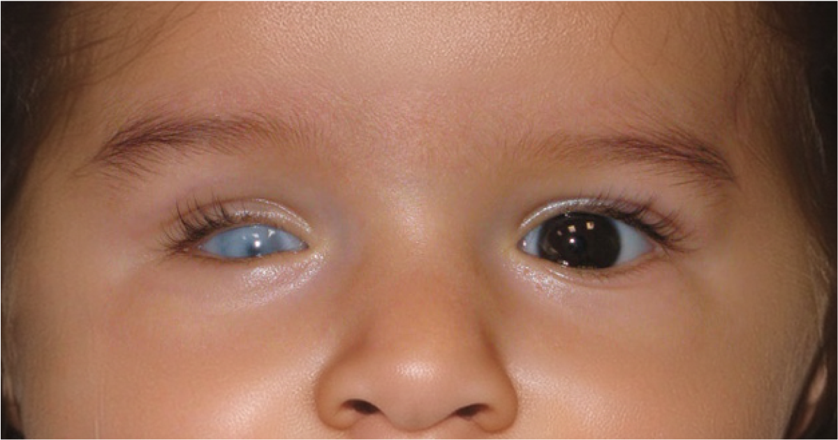
Learning when your baby’s eye is smaller than it is supposed to be is very alarming for parents. You may immediately question vision, facial growth, what the treatment will be, and your child’s outcome. While this is an atypical issue, in time and with coordinated care families may begin to understand what has put them in this situation and what support is available. Microphthalmia is a birth defect of the eye. This means the eye does not develop fully during pregnancy and is smaller than what is normal. What is put forth by microphthalmia is that it varies greatly. Some babies may have a barely noticeable smaller eye with good vision, while others may have large scale underdevelopment and serious visual impairment.
The diagnosis is a narrow scope of what a child will experience or how the issue will play out in terms of development. Vision potential is determined by what is present within the eye, health of the retina and optic nerve, also by associated eye issues and which eyes are affected. Thus an in depth evaluation by a pediatric ophthalmologist is very much so recommended.
What Is Microphthalmia?
Parents who are researching what is microphthalmia are often looking for a basic answer to a complex birth issue. Microphthalmia which is a term used when an eye does not fully develop before birth and as a result is abnormally small. It may affect the right eye, the left eye, or in some cases both eyes.
In some children the smaller eye does indeed include all typical eye elements which may just be reduced in size or did not form properly. In other cases we see the condition which goes along with cataract, coloboma, retinal issues, a small cornea, or underdevelopment of the optic nerve. Also these related issues often play a larger role in the degree of visual impairment than does eye size alone. The CDC reports that anophthalmia and microphthalmia which they study, develop during pregnancy and may appear by themselves, with other birth defects, or as part of a recognized syndrome.
Understanding the Microphthalmia Meaning
The medical microphthalmia meaning comes from words referring to an abnormally small eye. But that is not the full picture. Eye size may in fact be a symptom of more extensive issues with the development of the orbit, lid, and facial bones which we see in very severe cases.
A small appearance in one eye is not at all times a sign of that which is present. Issues of lid asymmetry, facial imbalance, drooping of the eye lid, or an eye which is positioned more deeply in the socket may also cause an eye to appear small. Diagnosis is through ophthalmic exam which in some cases will include imaging to accurately measure and assess the eye and socket.
Microphthalmia and Anophthalmia Are Different Conditions
Microphthalmia is at times put forward along with anophthalmia which it is a term for when the eye does not develop at all. In anophthalmia there is no eye structure that forms, but in microphthalmia while the tissue for the eye is present that which does develop is so under which the eye is very small. In severe cases of microphthalmia the eye may appear to be non existent during first examination. This is because the little eye tissue that is present is very small and may not be easily seen. Ultrasound or Magnetic Resonance Imaging may help to determine what eye structures are present.
| Condition | What develops before birth | Possible effect on vision | General care focus |
| Microphthalmia | A small, incompletely developed eye is present | Vision may range from useful sight to severe impairment | Preserving available vision, treating associated abnormalities, and supporting socket growth |
| Anophthalmia | One or both eyes do not form | There is no vision from the affected side | Supporting socket and facial development with conformers, prosthetic care, or selected surgery |
| Microcornea | The cornea is smaller than expected, while the entire eye may not be significantly small | Depends on other eye structures and associated conditions | Monitoring vision, refraction, eye pressure, and related abnormalities |
| Coloboma | A portion of eye tissue does not close or form completely | Depends on the location and extent of missing tissue | Visual assessment, correction of refractive error, and monitoring for complications |
How Does a Baby’s Eye Develop?
During early pregnancy the eyes begin to develop. A complex sequence of genetic signals which is very coordinated guides the formation of the eye, lens, retina, optic nerve, eyelids, and surrounding tissues. Should any part of this process be interrupted the eye may grow abnormally or some structures may not form at all.
As eye growth is a component of brain, face and other organ development, at diagnosis of microphthalmia we may note the health of other systems in the body. This is not to say every affected child also has another medical issue. It is true that many have a stand alone eye difference but a more in depth assessment will help identify associated issues that may improve with early intervention.
The issue may present as unilateral which is to say affecting one eye, or bilateral which is in both eyes. With unilateral microphthalmia a child may rely largely on the better seeing eye. Bilateral disease has a greater tendency to affect visual development, mobility, learning and communication but the degree of impact varies from child to child.
What Causes Microphthalmia?
In many of these early cases doctors are not able to determine a specific cause. Also it is important for parents to know that the diagnosis is not a result of what they may have done or not done during pregnancy. That which affects eye development is a bio complex issue and issues may come up even in very well managed pregnancies. We know that some causes are genetic which in turn affect the growth of the eye, also there are issues with the number or structure of chromosomes and in some cases the environment plays a role during pregnancy. Also at times a few factors may play together and in other cases we run out of explanations after due testing.
Genetic and Chromosomal Causes
Changes which affect many genes have been reported in microphthalmia. These genes play a role in the early development of the eyes and in some cases other organs also. A genetic change may be inherited from a parent, may follow different patterns of inheritance, or may appear for the first time in a child which has no family history of the issue.
MedlinePlus Genetics reports that many genes may play a role and that the condition which we are talking of has a tendency to present in an autosomal dominant or autosomal recessive or X linked pattern. Also it is important to note that just because a gene is associated with the condition does not mean we can predict the degree of eye change or visual impairment in a child. We may recommend a chromosome test or a more in depth genetic test when both eyes are affected, when microphthalmia is very severe, when other at birth physical differences are present, or when there is a family history. Also genetic counseling may help families out in understanding what a test result does mean, what it does not mean, and also what may be the issues related to future pregnancies.
Pregnancy-Related Environmental Factors
Certain infections, medicines which include some that affect fetal development, and nutritional deficiencies have been reported to cause anophthalmia and microphthalmia. As reported by reliable medical resources rubella infection, use of medicines like isotretinoin or thalidomide, and very serious vitamin A deficiency.
These associations should be put into perspective. We do not see that every pregnancy affected by an infection or medicine exposure will in turn present with an eye anomaly. Also we do not feel that parents should discontinue prescribed medication without first seeking medical advice. It is advised that any person pregnant or planning pregnancy should go over prescription medicines, over the counter products, and supplements with a qualified doctor. High dose vitamin A and vitamin A based medicines in fact may themselves be harmful during pregnancy. Also families should not try to prevent eye issues by taking large doses of supplements which have not been recommended by an obstetrician or some other qualified health care professional.
Isolated and Syndromic Microphthalmia
When that which is affected is the eye and does not have recognized problems in other parts of the body it may be reported as isolated microphthalmia. When it presents with issues in the brain, heart, hearing, kidneys, limbs, growth, hormones, or other systems it may be reported as syndromic. This distinction is not always able to be made at the time of the first visit. Some related features only present themselves after further testing or as a child grows. Thus pediatrics, genetic, neurological, hearing, development, or endocrine assessments may be recommended according to the child’s, at the time, health picture.
What Are the Main Microphthalmia Symptoms?
What is most noted is an eye which appears to be of a smaller size than the other eye or what is normal for the baby’s age. In severe cases the eye may be hard to see behind the eyelids. Also the eye socket or eyelid opening may appear smaller to which this condition may present in only one side.
Other microphthalmia symptoms and what to look for depends on which structures are affected. A baby may present with a small or cloudy cornea, an irregular pupil, a cataract, uncontrolled eye movements, misaligned eyes, delay in response to light, or reduced visual attention. Some abnormalities may go unnoticed by parents. A child can have a small retina, abnormal development of the optic nerve, refractive error, high eye pressure, or a coloboma within the eye which may not have a visible external sign. This is why a full dilated eye exam is important even when the baby seems to be doing fine.
| Sign noticed by parents or clinicians | What it may suggest | Recommended response |
| One eye or eyelid opening looks smaller | Underdevelopment of the eye, socket, eyelid, or surrounding tissues | Arrange a paediatric ophthalmology assessment rather than relying on appearance alone |
| The baby does not consistently follow faces or light | Reduced visual function in one or both eyes | Seek early assessment so usable vision and visual development can be evaluated |
| The eyes move repeatedly or do not align | Nystagmus, strabismus, or unequal visual input | Ask about refraction, amblyopia assessment, and appropriate visual rehabilitation |
| The pupil looks white, cloudy, unusually shaped, or different from the other eye | Cataract, coloboma, retinal disease, or another significant eye condition | Obtain prompt examination at an eye specialist hospital |
| Redness, swelling, discharge, pain, or sudden behavioural change | Irritation, infection, pressure-related problems, or another complication | Contact the treating ophthalmologist without waiting for the next routine visit |
How Is Microphthalmia Diagnosed?
Diagnosis may take place during pregnancy or after delivery. The timing depends on the severity of the condition, the quality and stage of prenatal imaging, and whether one or both eyes are affected.
Diagnosis During Pregnancy
A report from a detailed prenatal ultrasound may note an eye that is smaller than average or may note issues with the eye’s visibility. If a fetal eye abnormality is reported the obstetrician may refer to a specialist in fetal medicine. Also at times Fetal Magnetic Resonance Imaging may supply more info to the eyes, brain and surrounding structures.
Prenatal care does not at all times identify the degree of useful vision a baby will have. Also it may be hard to tell between severe microphthalmia and anophthalmia before birth. Thus some families may have to go through more evaluation post birth before they receive a clearer diagnosis and management plan.
Examination After Birth
At birth a pediatric ophthalmologist will check out the eyelids, eye opening, cornea, pupil, lens, retina, optic nerve, eye movements, visual responses. Also the doctor may compare the two eyes and note in the affected eye which structures for vision are present.
Ocular ultrasound is a tool which we use to measure the eye and also look at structures which do not present well through the pupil. We may turn to magnetic resonance imaging when it is required to study the optic nerves, eye sockets, brain or nearby tissues. The type of imaging that is done is based on what the baby requires and we are careful to avoid unnecessary radiation. A small eye may have large focusing errors which we correct early to improve the visual input to the brain. Also the better seeing eye must be evaluated carefully as at times associated issues may present in it.
General Medical and Genetic Assessment
A pediatrician may do a full physical exam which in some cases may reveal associated birth defects. For some cases in which history or exam results point to a more wide spread issue, hearing tests, development evaluation, heart or kidney study, hormone testing, neurology review, or other studies may be recommended.
Genetic testing is not identical for every child. The most suitable test depends on the pattern of eye findings, whether one or both eyes are involved, family history, and any associated medical concerns. Also a negative genetic result doesn’t rule out a genetic cause as we do not yet have tests for all developmental changes.
Can a Baby With Microphthalmia See?
Some babies can see through the affected eye, in some others’ that eye has very little or no vision at all. Eye size does not in itself tell the story. Vision depends on the health of the cornea and lens, the retina and optic nerve development, the connection between the eye and brain, and also the presence of treatable conditions. A small eye which has a healthy retina and optic nerve may still do fairly well. On the other hand an eye which has large scale retinal dysplasia, very poor optic nerve development, wide coloboma, or a dense congenital cataract may have very little visual prospect.
When there is a difference in vision between the two eyes the brain may start to use the eye with better vision more and at the same time reduce use of the weaker eye which in turn may cause amblyopia also known as lazy eye. We may use glasses, contact lenses, treatment for an obstructing cataract or closely supervised patching as a treatment when we think that the affected eye will be able to develop useful vision. Parents should not patch the better-seeing eye without instructions from a paediatric ophthalmologist. The duration and safety of patching depend on the baby’s age, visual potential, and the condition of both eyes.
Treatment for Microphthalmia
There is not a one size fits all treatment for any child. What we do is base it on the degree of eye development, available vision, the health of the other eye, size of the socket, associated health issues and the overall development of the child.
Main goals are to preserve what vision is present, promote balanced growth of the eye socket and adjacent facial structures, manage associated eye disease, support growth and also to present naturally when possible. The National Eye Institute’s guidance on anophthalmia and microphthalmia care report that they may use prosthetic devices, surgery, and medical treatment based on the child’s individual needs.
Correcting Vision and Preventing Amblyopia
When there is visual potential in the affected eye the ophthalmologist may put in glasses or in some cases a contact lens. We correct refractive error which in turn gives the developing brain a better quality visual input and may also reduce the risk of amblyopia.
For the a typical which may present itself between the two eyes patching or other amblyopia therapies may be used. This does require a great deal of monitoring as too much patching of the better eye can in fact interfere with its development.
That which is dependent on one eye is usually given well fitting glasses made from impact resistant lenses. As the child begins to move around protective wear is recommended to reduce the risk of accidental injury to the better seeing eye.
Treating Associated Eye Conditions
Some children need specific eye treatments for cataract, glaucoma, retinal problems, strabismus, corneal abnormalities, or other associated conditions. Surgery is a option when an issue which affects vision or which puts the eye’s health at risk is present but that is a call which is based on what we expect the benefit to be, visual prognosis, anesthetic issues and also what the complications may be.
Removing cataracts is a variable result in terms of recovery to normal vision if the retina or optic nerve are also under developed. Parents should get a frank discussion of what the surgery can do and what the follow up care will require.
Conformers and Eye-Socket Development
The eye is a factor in the growth of the surrounding socket and facial bones. If an eye is very small the affected socket may also grow less than the other. Thus early management of the socket may be recommended to support facial symmetry.
A conformer which is a smooth device is placed behind the eyelids. It is for maintaining or as the baby grows out to gradually expand the socket. As the child grows you may have to change the conformer for a larger size. Fit and adjustment should be done by professionals with experience in pediatric socket development. A conformer does not restore sight. It is to support the eyelids, socket and surrounding tissues. When vision is present in a small eye treatment must be planned out very carefully so that socket management does not in turn impair the child’s sight.
Ocular Prosthesis and Reconstructive Surgery
Once the socket has developed enough an ocularist may create a custom made prosthetic eye or cosmetic shell. The prosthesis is designed to fit the child’s socket and to improve appearance. It does not restore sight, but it may support facial growth and psychosocial health. As children grow very quickly a prosthesis may require to be adjusted, polished, or replaced. Parents should follow the ocularist’s instructions for insertion, removal, and cleaning which includes what products to use instead of home remedies or inappropriate products.
In some serious cases an oculoplastic surgeon may bring up the use of expanders, implants, grafts, or reconstructive procedures. Not all children with microphthalmia go right to surgery. The decision is very much a personal one which takes into account socket growth, eyelid structure, visual prospects, past treatment, and the family’s values. Clinical reports stress early input from a team of specialists which at the same time note that what works for one may not for another.
Low-Vision and Developmental Support
When there is great reduction in vision early support can help a child make use of what sight they have and also develop alternate ways of exploring the world. Low vision specialists may put forth high contrast objects, proper lighting, positioning strategies, visual stimulation, or age appropriate assistive tools.
In cases where visual impairment affects movement or learning physiotherapy, occupational therapy, orientation and mobility support, and early educational services may be useful. With limited vision a child can still develop communication, independence, social relationships, and academic skills when support is given at an early age and which also changes as the child grows.
Follow-Up Care and Long-Term Management
Microphthalmia for the most part is treated throughout childhood as an ongoing process as opposed to a one time correction. As the child grows out his or her care plan changes to include visual skills training, entry into the school system which may bring up different issues, and the child’s input in to what regards appearance and treatment. In follow up we see the ophthalmologist for evaluation of visual behavior, refractive error, amblyopia, eye alignment, cataract, retinal health, eye pressure, and the performance of the better doing eye. The oculoplastic surgeon or prosthodontist will look at socket growth, lid position, conformer fit, and prosthesis comfort.
Parents should report if they see persistent redness, swelling, pain, bleeding, discharge, repeated displacement of the prosthesis or conformer, or a sudden change in visual behavior. A prosthesis or conformer that no longer fits well should be brought to the attention of the health care team instead of forced into place. Also to be reevaluated are the child’s developmental and educational needs. A child which did well at home may now have trouble with reading, writing, in the classroom or with sports. Open communication between the family, health care team, therapists and teachers is key to addressing these changes as they happen.
Recovery and Expected Outlook
The word “recovery” can be misleading because microphthalmia is a developmental condition rather than a temporary illness. Treatment cannot make an incompletely developed eye anatomically normal. Instead we focus on what we can do to improve vision that is present, support growth of the socket, treat related conditions, and we work to get the child as active in day to day life as possible.
The outlook varies widely. A child with unilateral microphthalmia and normal vision in the other eye may do well enough to attend regular school which will include the use of proper eye protection. A child with bilateral visual impairment may require ongoing low vision services and educational adaptation.
After surgery or a conformer adjustment it is typical for a child to experience some swelling, watering, irritation or discharge which will vary by the procedure. Each family will get different aftercare info as recovery is not the same for all children and treatments. If you see increasing pain, fever, large scale swelling, bleeding or atypical discharge report that right away. Also emotional adjustment is a part of the long term care picture. Parents may feel grief, uncertainty, or guilt after diagnosis. Clear information, compassionate counselling, contact with appropriate support services, and realistic discussions about treatment can help families feel more confident.
Can Microphthalmia Be Prevented?
In many cases prevention is not possible which is due to the fact that the cause is not known or is a result of a random genetic mutation. A diagnosis is not a result of a parent’s care or in care during pregnancy. Pre and early pregnancy care may lower some of the avoidable risks. Women who are trying to get pregnant should talk to their doctor about their medications, also do not self medicate with retinoids or high dose supplements, get the rubella vaccine before getting pregnant, stay away from alcohol and other harmful substances, and report for all recommended antenatal care.
Rubella vaccination does not usually take place during pregnancy which is why it is best to review immunization status prior to conception. Also do not discontinue prescribed medication suddenly without first speaking to your health care provider as some maternal health issues left untreated may also present risks. In the case of past affected pregnancy, known genetic diagnosis, or history of congenital eye defects families may benefit from genetic counseling before trying again. Counseling will put forward info on inheritance and testing options which although very useful, in all cases does not guarantee what may or may not happen with a repeat pregnancy or the severity of any issue which may present itself.
When Should Parents Consult an Eye Specialist?
A newborn should have early assessment when it is seen that one eye is much smaller, the eyelids are out of the usual, the eyes do not react to light, the pupils look different, or the baby doesn’t show the expected visual attention. Also very important is the evaluation when a pupil appears white, the cornea is cloudy, the eye becomes red or painful, the eyelids are greatly swollen, or the child suddenly stops using vision as before. These signs may indicate a related health issue which requires prompt treatment.
Families should choose an eye hospital or eye specialist hospital with access to paediatric ophthalmology service as well as at the ready oculoplastic, genetic, pediatric, low vision, and ocular prosthetic services. Coordinated care is very much a value added component to the care we provide for the fact that a child’s visual, medical, developmental and cosmetic needs may change over time.
Supporting Your Child With Confidence
A diagnosis of microphthalmia may be very tough to hear but it is important to know that it does not define who your child is, what they can do or what their future holds. Some children just need routine vision checks and protective glasses, in other cases a mix of visual rehabilitation, conformer therapy, prosthetic care, surgery, and developmental support may be required.
Parents play a key role in taking their child to all follow up appointments, in following through with amblyopia and prosthesis care instructions, in protecting the better seeing eye, in noting any changes in visual behavior, and in reporting back to the school and health care team. While early evaluation doesn’t determine the visual outcome, it does allow doctors to identify issues which may be treated as the visual system and facial structures are still developing. For best results consult a qualified pediatric ophthalmologist at Vasan Eye Care or a trusted eye hospital which will do a full assessment.
Frequently Asked Questions
Small eyes can be a result of incomplete eye development, genetic or chromosomal changes, certain pregnancy related infections, or from exposure to some drugs. In many babies a cause is not determined. An ophthalmologist should confirm that what you see is microphthalmia and not some other eye or lid issue.
What is microphthalmia small eye syndrome is a term used by families to describe a birth defect which includes the size of one or both eyes to be below the normal range and also underdeveloped. It may present by itself or with other eye, physical or medical issues which require a coordinated evaluation.
Yes, some babies with microphthalmia retain useful vision, while others have severe impairment. Vision depends on the retina, the optic nerve, the lens, the cornea and also the connections to the brain which isn’t related to eye size alone. A pediatric ophthalmologist has to look at each eye separately.
That they may have microphthalmia, a small cornea, lid differences, facial asymmetry or some other development issue. Appearance alone does not determine cause or visual prognosis. Parents should take the baby in for an evaluation at a qualified eye specialist hospital for an accurate diagnosis and personalized care.
References
- Centers for Disease Control and Prevention: Anophthalmia and Microphthalmia
https://www.cdc.gov/birth-defects/about/anophthalmia-microphthalmia.html - MedlinePlus Genetics: Anophthalmia and Microphthalmia
https://medlineplus.gov/genetics/condition/anophthalmia-microphthalmia - National Eye Institute: Anophthalmia and Microphthalmia
https://www.nei.nih.gov/eye-health-information/eye-conditions-and-diseases/anophthalmia-and-microphthalmia - PubMed Central: Management of Anophthalmia, Microphthalmia and Coloboma in Newborns
https://pmc.ncbi.nlm.nih.gov/articles/PMC11881466