
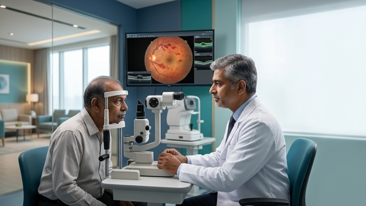
BRVO causes a small vein in the retina to block. Blood or fluid may leak into the retina which in turn causes macular edema. Timely branch retinal vein occlusion treatment is key in managing these issues and in preserving vision.
What Is Branch Retinal Vein Occlusion?
A retinal artery often develops over a branch vein which in turn causes compression. According to the National Eye Institute report on retinal vein occlusion they note that Branch retinal vein occlusion treatment (BRVO) is of the smaller retinal veins and not the large retinal vein. Treatment for branch retinal vein occlusion is based on the site of the blockage, macular involvement and degree of vision loss.
A branch vein occlusion may present with bleeding, swelling or diminished circulation, thus branch retinal vein occlusion treatment follows that of a full retinal exam.
Branch Retinal Vein Occlusion Symptoms
Common branch retinal vein occlusion symptoms are sudden painless vision blurring, distorted central vision or a dark spot in part of the visual field. Retinal vein blockage symptoms present more when the macula is swollen.
| Retinal change | Possible visual effect |
| Macular edema | Blurred or distorted central vision |
| Retinal bleeding | Hazy vision or a dark patch |
| Reduced circulation | Partial visual-field change |
Sudden vision loss should be quickly evaluated. Evaluation will determine which branch retinal vein occlusion treatment or which course of close monitoring is appropriate.
Risk Factors for BRVO
A branch vein occlusion is related to vascular risk factors. MedlinePlus reports that high blood pressure, diabetes and atherosclerosis are associated with retinal vein occlusion which also notes that glaucoma and increasing age are recognized associations. These points are important in the treatment of branch retinal vein occlusion treatment.
A patient that has brvo eye may also require management of blood pressure, blood sugar and other vascular risks along with branch retinal vein occlusion treatment.
How Is BRVO Diagnosed?
Diagnosis usually performs visual acuity tests and dilated retinal exams. We also at Vasan Eye Care may do a retina test like optical coherence tomography (OCT) which measures macular swelling. For more in depth info on leakage or retinal blood flow we may use fluorescein angiography. That which we see guides branch retinal vein occlusion treatment.
| Test | Why it may be used |
| Dilated retinal examination | Checks bleeding, swelling and the affected area |
| OCT | Measures macular edema and monitors change |
| Fluorescein angiography | Assesses leakage and retinal circulation |
Repeat imaging helps monitor branch retinal vein occlusion treatment.
Branch Retinal Vein Occlusion Treatment Options
The primary goal of branch retinal vein occlusion treatment is to prevent complications which affect vision, mainly macular edema. If vision is preserved and swelling is minimal, watchful waiting may be recommended.
When macular edema affects vision, at Vasan Eye Care we see that anti-VEGF agents are very much used to reduce leakage and swelling. This is a key part of our brvo treatment. In some cases we may also look at corticosteroid therapy. We also at Vasan Eye Care use Retinal laser photocoagulation which is useful in certain situations. At NCBI we find that anti-VEGF therapy, corticosteroids and lasers are recognized approaches for BRVO complications. Branch retinal vein occlusion treatment we tailor to what we see in the retina and how the patient responds to treatment. What we do in the management of BRVO is also an ongoing process of review.
Regular follow up is important because branch retinal vein occlusion treatment may require repeat assessment. OCT is used to track macular edema, also blood pressure and sugar levels play a role in care. Branch retinal vein occlusion treatment may change as the eye does in response.
Can Vision Improve After BRVO?
Vision results are variable. In some cases eyes improve as the swelling goes down, in other cases we seem to be dealing with a more permanent form of branch retinal vein occlusion treatment. Macular involvement and also reduced circulation play a role in how well one does.
If retinal vein blockage symptoms present in an advanced stage, or there are sudden new floaters or severe vision loss, it is advised to get in for an urgent examination. At Vasan Eye Care we provide retina services Vasan Eye Care’s retina services for evaluation, imaging and treatment planning. Timely care is key to our approach in branch retinal vein occlusion treatment which is directed at the vision affected complications.
Frequently Asked Questions
1. What is the usual treatment for BRVO?
Branch retinal vein occlusion will also at times include an option of watchful waiting, delivery of anti-VEGF agents, administration of steroids if which is appropriate, also laser treatment as well which will be based on the results of retinal examination.
2. Is BRVO an emergency?
Sudden vision loss which requires immediate assessment, also when BRVO symptoms present out of the blue.
3. Can BRVO improve without treatment?
Some cases do see improvement but in other cases complications may persist. An ophthalmologist has to determine if branch retinal vein occlusion treatment or just monitoring is appropriate. In most cases of branch retinal vein occlusion treatment is not required.
4. What causes vision loss in BRVO?
Macular edema is a frequent cause of reduced vision and a focus of brvo treatment.
5. Can BRVO happen again?
Yes. Recurrence or occlusion of the other eye is an issue which presents when vascular risk factors are not controlled. Ongoing follow up is supported for branch retinal vein occlusion treatment and risk factor management.
References
- Central Retinal Vein Occlusion (CRVO) – National Eye Institute (NEI)
https://www.nei.nih.gov/eye-health-information/eye-conditions-and-diseases/central-retinal-vein-occlusion-crvo National Eye Institute - Retinal Vein Occlusion – MedlinePlus Medical Encyclopedia
https://medlineplus.gov/ency/article/007330.htm MedlinePlus - Branch Retinal Vein Occlusion – StatPearls, NCBI Bookshelf
https://www.ncbi.nlm.nih.gov/books/NBK535370/ NCBI