
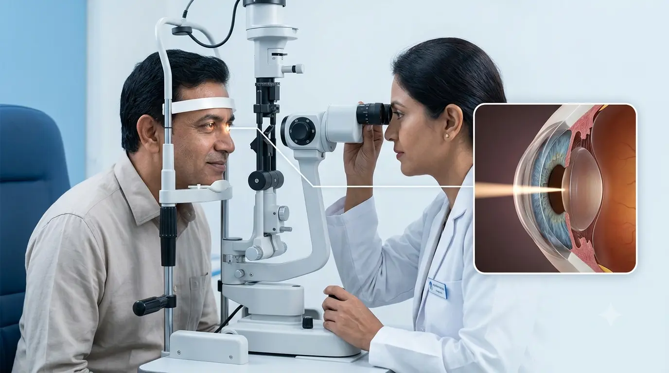
An eye exam is far more in depth than performing which letters are present on a visual chart. Some ocular diseases start in a very incipient stage of change that can only be detected with magnification. The slit lamp is used by the ophthalmologist to see these initial changes better and also to assess various eye structures.
A slit lamp eye exam uses a special microscope in association with a bright adjustable beam of light. In this exam the doctor is able to study structures which include the eyelids, conjunctiva, cornea, iris, anterior chamber and lens in great detail. Also with the use of additional lenses and pupil dilation the doctor may look at the vitreous, retina and optic nerve.
The American Academy of Ophthalmology reports that the slit lamp is a very useful tool which gives ophthalmologists a close up view of the eye’s components.
What Is a Slit Lamp?
A slit lamp is a type of binocular microscope that is paired with a very bright light source. As opposed to a normal torch the device is able to put out a thin and intense beam of light. Also the health care professional is able to change the width, height, brightness and angle of this beam while doing an eye exam.
During the slit lamp exam the patient places their chin and forehead into supports which are a part of the instrument. The ophthalmologist which is on the other side sits at the microscope which is also the source of light for each eye.
The right mix of lighting and magnification which we get from the slit lamp gives the doctor a in depth 3D view of many eye structures. Thus the slit lamp test is a very important part of a full medical eye exam.
Why Is a Slit Lamp Examination Performed?
The primary purpose of a slit lamp eye exam is to look for issues which may not present in a routine vision test. It is possible for a person to have 20/20 vision and at the same time have early stages of change in the cornea, lens, tear film or other internal structures. With a thorough examination, an ophthalmologist is able to see these changes and to decide which if any further tests or treatment is required.
The slit lamp uses range from routine eye exam to diagnosis and treatment of specific eye diseases. Also it is a tool which doctors use to evaluate eye injuries, inflammation, infections, dry eye symptoms and changes related to age.
For persons which have an eye condition, through repeated exams the ophthalmologist is able to see how the condition progresses over time and to assess the results of the treatment.
Which Parts of the Eye Can Be Examined?
During a slit lamp examination which structures are looked at may depend on what the patient reports, past health issues, if we put in extra lenses or use pupil dilating drops.
| Eye Structure | What the Ophthalmologist May Examine |
| Eyelids and eyelashes | Inflammation, swelling, abnormal lashes and eyelid problems |
| Conjunctiva | Redness, irritation and inflammation |
| Cornea | Scratches, infections, ulcers, swelling and scars |
| Anterior chamber | Inflammatory cells, abnormal depth and other changes |
| Iris | Shape, inflammation and abnormalities |
| Lens | Cataract and other lens changes |
| Vitreous | Opacities or abnormalities when clinically indicated |
| Retina and optic nerve | Retinal or optic nerve changes using additional lenses and, when needed, pupil dilation |
This ability to look at a variety of structures is what we see in slit lamp examination uses which has application across many areas of ophthalmology. MedlinePlus reports that a slit-lamp exam may also help to identify conditions which affect the cornea, lens, retina and other eye elements.
What Happens During a Slit Lamp Test?
A slit lamp test is generally straightforward and does not require an incision, injection or surgical procedure.
First of all you will sit in front of the equipment, then the ophthalmologist or eye care professional will get you to put your chin into the chin rest and your forehead against the frame. For best results keep your head very still as it allows the doctor to use the microscope precisely.
The doctor will pass a thin beam of light into one eye at a time which will appear very bright but of a short duration. We may change the light’s angle and width in order to see different inner layers and structures. You may be asked to move your eye by looking in different directions which will in turn better allow certain areas to be seen. Also in some cases we will use eye drops during the slit lamp exam. The type of drop will depend on what the doctor is looking for.
Why Is Fluorescein Dye Sometimes Used?
An ophthalmologist may at times use fluorescein in a slit lamp eye exam in which the surface of the eye requires close evaluation. Fluorescein is a diagnostic dye in which the corneal surface has had damage or disruption does its best to show up. When viewed under blue light areas which are broken like in the case of a corneal abrasion or certain surface defects stand out more.
According to MedlinePlus, fluorescein staining can help detect foreign bodies and damage to the cornea.
The dye usually disappears naturally with blinking and tears.
Can Eye Pressure Be Checked Using a Slit Lamp?
Yes. A technique called applanation tonometry may be performed with a slit lamp to measure intraocular pressure.
Before this test, numbing drops are generally placed in the eye. A small measuring device then briefly touches the surface of the cornea. The measurement helps ophthalmologists assess eye pressure, which forms an important part of glaucoma evaluation and monitoring.
According to MedlinePlus fluorescein staining is a test which may be performed to identify foreign bodies and damage to the cornea. The dye in most cases will go away naturally with blinking and tears.
What Conditions Can a Slit Lamp Examination Detect?
A slit lamp examination is a tool which does not diagnose all eye conditions. What it does is present results which may be indicative of a problem which in turn will guide which tests are run.
| Condition or Problem | Possible Slit-Lamp Findings |
| Cataract | Clouding or changes within the natural lens |
| Dry eye | Tear-film abnormalities and ocular-surface changes |
| Corneal abrasion | Surface defect, often highlighted using fluorescein |
| Corneal infection or ulcer | Inflammation or abnormal areas in the cornea |
| Uveitis | Signs of inflammation within the front portion of the eye |
| Eye injury | Foreign material, surface damage or structural changes |
| Conjunctivitis | Redness, discharge and conjunctival inflammation |
| Retinal or optic nerve disorders | Changes visible with additional examination lenses and appropriate dilation |
The examination results should be put in the context of symptoms, visual acuity, medical history and also any additional tests which the ophthalmologist suggests. MedlinePlus reports that cataracts, corneal injury, dry eye, uveitis and also a number of retinal disorders are among the conditions which may be brought to light during slit-lamp assessment.
Will My Pupils Be Dilated?
Not for all slit lamp eye exam does dilation take place. The front parts of the eye may in fact be looked at without dilating the pupils. But if it is the retina, the optic nerve or other back of the eye structures which the ophthalmologist has a look at, we may use dilating drops. Dilated pupils may for a while cause close up vision to go blurry and also increase sensitivity to bright light. Patients may find that they are more at ease wearing sunglasses after a dilated exam.
Is a Slit Lamp Examination Painful?
A very common feature of a routine slit lamp test is that it is pain free. During the basic exam no items enter the eye. The bright light may at first be uncomfortable for a few moments which is also more likely if you are very light sensitive. Should other procedures which include but are not limited to measurement of eye pressure become necessary, numbing drops will be used at appropriate times. It is always best to report to the ophthalmologist if you do experience great discomfort, sudden eye pain or any other odd symptoms during an exam.
Who May Need a Slit Lamp Eye Exam?
A slit lamp exam forms part of many comprehensive ophthalmic evaluations. Also it may be very useful in case of a patient which reports persistent redness, eye pain, blurry vision, light sensitivity, excessive tearing, dry eyes issues or that had a reported eye injury.
People who have cataracts, corneal disorders, glaucoma, diabetes related eye issues or previous eye surgery also may have slit-lamp exams as a part of follow up care. While routine eye exams may be recommended at which the vision seems fine they are still very much so a good idea because many eye diseases can develop before any symptoms present themselves.
At Vasan Eye Care an eye specialist doctor will assess your symptoms and determine which tests are right for your eye health.
Conclusion
The slit lamp is also the very useful tool which we use in ophthalmic exam. It magnifies the area of the eye which at the same time is also subjecting it to a very fine beam of light which in turn allows the ophthalmologist to look at the structures which range from the eyelids and cornea to the lens and with the help of certain techniques also into the deeper parts of the eye.
A slit lamp eye exam which is also used in glaucoma, retinal and follow up exams is a tool which reports on cataracts, corneal issues, inflammation, dry eye changes, injuries and also many other abnormalities.
If you experience persistent redness, pain, blurred vision, light sensitivity or any other unusual eye symptoms do take into your care an eye hospital instead of waiting for the symptoms to worsen. At Vasan Eye Care our ophthalmologist will do the appropriate exam and we will recommend further testing or treatment based on our findings.
Frequently Asked Questions
1. How long does a slit lamp examination take?
A short time is what a basic slit lamp examination takes, though the full eye appointment may last a little longer. What we see depends on the structures which are being looked at also what other procedures if any are carried out such as dilation, fluorescein staining, tonometry or retinal evaluation.
2. Can a slit lamp detect cataracts?
Yes. One of the important slit lamp uses is that it allows for a close look at the natural lens of the eye. What we see through this tool is which cataracts may be present and how they are changing. Also other tests may be done to determine what degree the cataract is playing a role in the loss of vision.
3. Can a slit lamp examination detect glaucoma?
A slit lamp eye exam is a tool in the assessment of glaucoma, but it does not by itself diagnose the condition. Eye pressure measurement, inspection of the optic nerve and tests like visual field testing or optical coherence tomography may also be required based on what the ophthalmologist sees.
4. Is bullseye lamp examination the same as a slit lamp examination?
No. The term bullseye lamp examination is a reference to what we use in traditional illumination for ENT exams which is to be distinguished from ophthalmic slit lamp biomicroscopy. A slit lamp is a specialized microscope which we use for in depth study of the eye structures.
5. Can children undergo a slit lamp examination?
Yes. Children at times may go through a slit lamp test as per the ophthalmologist’s assessment. Also which the exam will go through may in part depend on the child’s age and ability to stay fairly still while the doctor does the eye exam.