
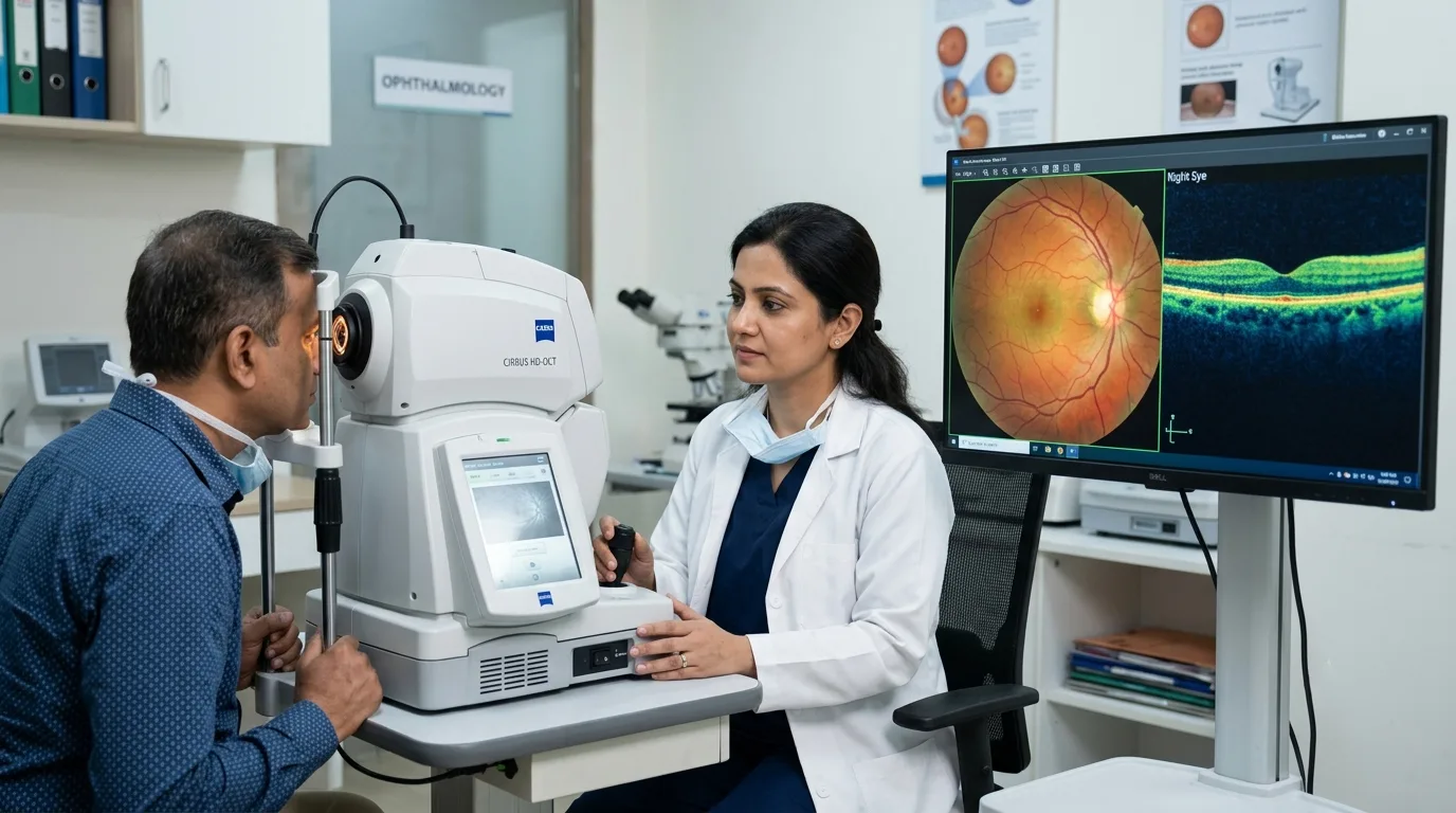
A healthy retina is at the core of clear vision. In the back of the eye is a thin light sensitive layer which also receives light and turns it into signals that go to the brain. At times changes in the retina may happen out of noticeability, without pain or any early stage vision issues. This is why we make use of retina scan and in depth retinal exams as a part of preventative and diagnostic eye care.
A retina scan which is used by eye doctors to get a much better in depth look at the back of the eye than what is usually possible with a routine vision test. As per your symptoms, health history, age, and risk factors, your ophthalmologist may suggest an OCT scan, retinal photography, dilated eye exam or another type of special retina test.
Understanding which tests will be done can make that upcoming retina check up feel much easier. This guide goes over the different kinds of retinal tests we have, what to expect during the procedure, how results are presented and when further investigation may be required.
What Is a Retina Test?
If you have been asking what is retina test is, what I can tell you is that we do not have a single definition for it. Instead it is a term that is used for a variety of tests and imaging procedures which we use to study the retina, macula, retinal blood vessels and other structures at the back of the eye.
For instance in a routine retinal examination a patient may see an ophthalmologist who will use special lenses and an ophthalmoscope or slit lamp to look through the pupil. In ophthalmoscopy the doctor is able to look at the retina, the optic disc, the choroid and the retinal blood vessels.
Modern there is a trend towards very detailed retinal imaging. For example, Optical Coherence Tomography, also known as OCT, uses light waves to produce in depth cross sectional images of retinal tissue. Also from the National Eye Institute we learn that OCT is a tool which eye doctors use to study retinal structures and to see changes like retinal or macular swelling. Thus when a doctor prescribes a retina scan the test is determined by what they are trying to research.
Why Is a Retina Scan Done?
A retina scan is a service which may be included in a routine eye screening, but we pay special attention to it when a person reports symptoms or has health issues which put them at high risk for retinal disease. Retinal changes are seen in diabetic retinopathy, age related macular degeneration, retinal vein occlusion, retinal tears, retinal detachment and also in many inherited retinal disorders. Also with the use of OCT imaging we are able to see and report on fluid or retinal swelling in the different layers of the retina.
People which do have diabetes require special care as diabetic retinopathy may present before we see any changes in vision. Diabetic eye screening is in to identify these early changes which at this point may be treated or monitored to help protect vision. An ophthalmologist may thus recommend a retina check up even when your eyesight seems normal.
Types of Retina Tests and Examinations
Different retinal tests provide different information. Your doctor may use one test or combine several investigations to obtain a complete picture of your eye health.
| Retina Test | What It Checks | Common Uses |
| Dilated retinal examination | Retina, optic nerve, macula and blood vessels | Routine retinal evaluation, diabetes, retinal tears and other eye diseases |
| OCT retina scan | Detailed layers and thickness of the retina | Macular swelling, diabetic changes, macular degeneration and retinal disorders |
| Fundus photography | Photographic image of the back of the eye | Monitoring retinal changes and diabetic eye screening |
| Fluorescein angiography | Blood flow and leakage from retinal blood vessels | Diabetic retinopathy, abnormal blood vessels and macular disorders |
| Fundus autofluorescence | Changes within retinal tissues | Selected inherited and degenerative retinal diseases |
| Electroretinography (ERG) | Electrical response of the retina to light | Inherited retinal conditions and retinal function assessment |
OCT Retina Scan
Optical coherence tomography is the primary tool which we use for retina scan. The patient will rest their head on the OCT machine and look at a target as the equipment takes images of the retina.
No at usual time that an instrument makes contact with the eye during routine OCT scan. The test produces detailed views of the retinal layers which in turn help the eye care professional to identify areas of swelling, thinning or other structural changes.
Dilated Retinal Eye Exam
During a retinal eye exam, it’s common for the eye drops that will dilate your pupils, which means the pupils get larger. As soon as they are dilated the ophthalmologist can have a more complete picture of the retina. At this point the doctor is also able to use indirect ophthalmoscopy or a slit lamp with special lenses. From this dilated retina the doctor can identify retinal tears, diabetic damage, abnormality in the optic nerve and many other eye issues.
Fundus Photography
Fundus photography produces images of the retina. These photos allow doctors to assess the state of retinal blood vessels, the optic disc and other structures. As photos can be saved and compared over time this type of retina test is very useful when doctors have to track changes between appointments. Also retinal photography is very much used in diabetes related eye screening.
Fluorescein Angiography
Fluorescein angiography is a test which shows retinal blood flow. We inject a fluorescent dye into a vein usually in the arm. As the dye goes into the blood stream and gets to the eye we take special photos. The test is able to identify leaking, blocked or abnormal retinal blood vessels. It is used in the diagnosis of conditions like diabetic retinopathy or macular edema.
Retinal Function Test
A structural retina scan gives us a picture of what the retina looks like, but at times an ophthalmologist requires information on its function.
An electroretinogram, or ERG, is a specialised retinal function test that measures the retina’s response to light. It is especially useful in the case of inherited and functional retinal diseases. Also the National Eye Institute reports that ERG is used in the diagnosis of conditions like retinitis pigmentosa.
What Happens During a Retina Check Up?
During your retina check up in we will start with a discussion of your symptoms and health history. Report to your ophthalmologist on diabetes, high blood pressure, past eye surgery, eye injuries, current medications and also any history of retinal or eye disease in your family.
Your vision will first be evaluated using a standard eye chart. Also we may do tests which check eye pressure or other components of the eye before we begin the retinal examination. If dilation is needed, we will put in drops for your pupils to open up. This may cause a brief stinging sensation. Once the pupils are dilated your ophthalmologist is able to see a larger area of the retina which we may also photograph.
For an OCT retina scan what you do is step up to the machine, put your chin and forehead in the proper place and look at the fixation target which takes the images. The process is usually very quick and non-invasive.
The time for a retina test is variable based on what is required for the exam. A basic exam may take only a few minutes, but dilation and special imaging may stretch the length of the appointment.
Is a Retina Scan Painful?
Most retina scan are pain free. During a retinal eye exam bright lights may at first feel out of comfort. Also you may experience a stinging sensation from the dilating drops which is followed by blurry vision and light sensitivity.
MedlinePlus reports that it is not uncommon for vision to be blurred for a few hours after eye dilation. We recommend that patients which still have blurred vision be given sunglasses and to arrange for a ride home. For tests which include injections of special dyes or the use of electrodes like in Fluorescein angiography or ERG different prep and feelings may apply. Your ophthalmologist will go over what to expect before we do the procedure.
Understanding Retina Scan Results
There isn’t a single “normal value” that applies to all retina scan. What you see depends on the test used and which structures are looked at. Your ophthalmologist will interpret the retinal images or report from the scan in the context of your symptoms, vision, age, medical history and past test results.
| Possible Finding | What It May Indicate |
| Normal retinal appearance | No significant abnormality detected on the examination |
| Retinal swelling or fluid | May occur with diabetic macular oedema, retinal vascular conditions or inflammation |
| Abnormal retinal blood vessels | May require investigation for diabetic or vascular retinal disease |
| Retinal thinning or structural changes | Can occur in several retinal or macular conditions |
| Tear, hole or detachment | Requires prompt ophthalmic evaluation and sometimes urgent treatment |
| Abnormal retinal electrical response | May suggest a functional or inherited retinal disorder |
In some cases of abnormal retinal examination it does not mean that severe vision loss will take place. Some issues may just require close monitoring, while others may need more in depth imaging or treatment. The ophthalmologist will go over what this all means for your particular situation.
Retina Test for Diabetes
A regular diabetes eye test is a key part of diabetes care which may in fact present without any noticeable symptoms at diagnosis. High blood sugar over time damages small retinal blood vessels which in turn cause leakage, swelling or the growth of abnormal new blood vessels.
The National Eye Institute reports that a dilated eye exam is a key tool in the diagnosis of diabetic retinopathy. A diabetes related retina scan may include retinal photography, OCT imaging or a detailed dilated exam which will vary based on the patient’s health reports.
The National Eye Institute says that for the most part people with diabetes should have a dilated eye exam once a year although the right interval in each case is to be determined by the eye care provider based on individual risk and past results. Also do not put off your diabetes eye test because your vision seems to be fine at present.
Who Should Consider a Retina Check Up?
A retina check up is especially important for people with diabetes, high blood pressure or an existing retinal condition. Also your ophthalmologist may recommend retinal imaging if you develop unexplained blurred or distorted vision, persistent visual changes or when changes are reported in a routine eye exam.
People which have had retinal disease in the past may require repeat retinal examination appointments as we compare the present with the past. The frequency of these exams should be a very personal matter as opposed to a set age based schedule. An ophthalmologist will determine how often you should have a retinal exam after they review your full eye and health history.
What Is the Retina Test Cost?
The retina test cost does not the same for all patients out that what we term as retina testing may consist of a series of different interventions. A routine dilated retinal exam is very different from something like OCT imaging, fluorescein angiography which is performed under general anesthesia or an in depth retinal function study like ERG. Also the total retina test cost may include the type and number of the tests ordered, the facility be it a hospital or a clinic, city which you are in, specialist’s consultation fees also play a role and at time more imaging may be required.
Rather instead of choosing a test which is the cheapest we should have an ophthalmologist determine what is medically appropriate first. Patients also may ask the hospital what the current charges are for the recommended procedure.
When Should You Seek Urgent Retinal Care?
Some retinal symptoms should not wait for a routine appointment.
A sudden onset of many floaters which are new, or flashes of light that repeat, or a dark curtain or shadow which moves in your field of vision can be signs of a retinal tear or retinal detachment. The National Eye Institute says to seek immediate ophthalmic care when these symptoms present themselves as retinal detachment can be a medical emergency.
Sudden loss of vision also requires prompt medical assessment.
Conclusion
A retina scan is a tool which ophthalmologists use to obtain info on retinal health and structure. As per what is required, a full retinal work up may include a dilated retinal examination, OCT imaging, retinal photography, angiography or a specialized retinal function test.
These tests are of great value in the management of diabetes, unexplained vision changes, or known retinal conditions. Also some retinal disorders may present with no clear symptoms at which point appropriate eye screening may identify a problem that may not yet be causing large scale visual issues.
If you have diabetes, notice any new vision issues or have been told to get a retina test, see the ophthalmologists at Vasan Eye Care which will be tailoring their evaluation to your specific eye health. Go to the emergency room for sudden flashes, a large increase in floaters, a curtain like shadow or sudden loss of vision.
Frequently Ask Questions
- What is a retina scan used for?
A retina scan produces in depth information on the structures at the back of the eye. As per the imaging method used it also helps ophthalmologists to evaluate the retina, macula and retinal blood vessels and to monitor changes related to diabetes, macular disorders or other retinal conditions.
- What is retina test and how is it performed?
People that are searching for what is retina test is to which we can answer that they are either referring to retinal imaging or a dilated exam. Your doctor may look at the retina during a dilated pupil or may use in office technologies like OCT or fundus photography. The exact procedure will depend on the health issue which brought you in.
- How long does a retina scan take?
The retina scan itself only takes a few minutes. But the full appointment may stretch out when there is dilating of the pupils, vision tests, consultation with the specialist, or if there is to the do additional retinal tests.
- Is dilation necessary for every retina test?
No. Some imaging tools do have the ability to take in high quality retinal images without the need for dilation. But we may see better and more in depth results when the ophthalmologist requires a wide view during a retinal eye exam. Your doctor will determine if dilation is a requirement.
- Can a retina scan detect diabetic retinopathy?
Yes. Retinal exam and imaging is a tool to identify changes in diabetic retinopathy. A diabetes eye test may include retinal photography, dilated exam and OCT based on the clinical picture. Early in the disease retinal changes may happen before a person is aware of vision issues.
- Is a retinal function test the same as an OCT scan?
No. OCT reports in detail on retinal structure of layers. As for the retinal function test we can assess with electroretinography that reports back how the retina does in terms of electricity in response to light. Doctors use these for various purposes and may at times suggest both.