
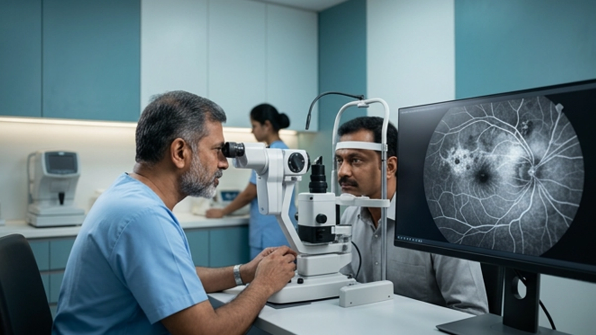
A procedure which uses fluorescein angiography eye test to image the blood vessels in the back of the eye is what we do in a fluorescein angiography eye test. We use it to see out for leakage, blocked circulation, abnormal new vessels and macular changes. Also we turn to this when a regular exam or OCT does not give us a full picture of a vascular issue.
What Is a Fluorescein Angiography Eye Test?
Fluorescein angiography which is also known as fundus fluorescein angiography (FFA) is a test that looks at circulation in the retina and choroid. Sodium fluorescein is injected into a vein usually in the arm. A retinal camera takes timed photos as the dye makes its way to the eye.
At the NCBI clinical review of fluorescein angiography it is reported that the ffa test eye evaluates retinal and choroidal circulation.
Why Is the Fluorescein Angiography Eye Test Recommended?
An ophthalmologist may recommend a fluorescein angiography eye test for diabetic retinopathy, macular edema, retinal vein occlusion or central serous retinopathy. Also it can determine poor blood flow and abnormal new vessels.
fluorescein angiography retina may play a role in treatment planning. This retinal angiography test is to identify capillary non-perfusion or a focal leakage point.
| Condition | What FFA can help show |
| Diabetic retinopathy | Leakage, non-perfusion and new vessels |
| Macular oedema | Pattern and extent of leakage |
| Retinal vein occlusion | Ischaemia and neovascularisation |
| Central serous retinopathy | Site and pattern of leakage |
How Is the Fluorescein Angiography Eye Test Performed?
Before performing the fluorescein angiography eye test the clinical team goes over past dye reactions, allergies, pregnancy and relevant medical history. We may use dilating drops for better retinal photos. Also included in a full eye test may be an OCT.
During the course of an eye angiography test, work up we place an IV in the hand or arm. Fluorescein is given in through the IV and we take photos which we do for several minutes as the dye goes through the retinal system. There may be temporary skin color change to yellow orange and also bright colored yellow urine.
What Do Fluorescein Angiography Results Mean?
A fluorescein angiography eye test is not a black and white issue. What we do is compare early and late images for signs of leakage, delayed filling, and blocked fluorescence. If the bright areas grow over time that is a sign of leakage.
Also we see dark areas as a sign of blocked fluorescence or reduced blood flow. Results are noted in context of the total exam and OCT.
| Angiography pattern | Possible interpretation |
| Increasing bright area | Dye leakage |
| Persistent dark area | Blocked fluorescence or poor perfusion |
| Delayed filling | Impaired circulation |
| Abnormal new vessels | Neovascularisation |
Fluorescein Angiography Side Effects and Precautions
Most of the time fluorescein angiography side effects are temporary. Nausea, vomiting, itching, hives or injection site discomfort may present. Also it is common for urine to turn very yellow for a short while after the fluorescein angiography eye test.
Anyone who has had a past serious fluorescein reaction should report it to the ophthalmologist before testing. Pregnancy in most cases is a relative contraindication. Breathing difficulty, facial swelling, extensive hives, fainting or chest issues require immediate medical attention.
Fluorescein Angiography vs OCT and OCTA
Most of what we see in fluorescein angiography side effects is of a temporary nature. That which we get from the fluorescent angiography eye test and OCT is different. OCT does what is to see retinal structure and fluid, on the other hand OCT angiography does what is to map blood flow out of intravenous dye. FFA is still a useful tool when we need to see active leakage and dynamic filling.
For retinal detachment which is a given the ophthalmologist will choose FFA, OCT, OCTA, ultrasound or a mix of them as per clinical presentation.
Eye Care at Vasan Eye Care
Vasan Eye Care ophthalmologists may also order a fluorescein angiography procedure if the which is to include blood vessel assessment is in the best interest of the patient’s diagnosis or treatment. May be performed at the time of the dilated exam or along with other retinal imaging.
Persistent or sudden changes in vision should be paid attention to. At Vasan Eye Care we have appointment times available for eye exam and testing.
Related Retina Tests and Eye Conditions
Fluorescein angiography helps eye care professionals examine blood flow in the retina and identify certain retinal abnormalities. To learn more about related diagnostic procedures, explore this guide to the retinopathy test and understand how doctors evaluate retinal health. You can also read about retinal vein occlusion treatment and the available options for managing this condition. Understanding image formation on retina can help you learn how the eye processes visual information. If a retinal vein blockage affects the central retinal vein, find out more about central retinal vein occlusion treatment. An eye specialist may also perform a slit lamp exam to examine the structures at the front of the eye. For further information about retinal assessments, explore the retina test and consult an eye care professional about the appropriate examination for your needs.
Frequently Asked Questions
1. Is a fluorescein angiography eye test painful?
The issue of pain is that it shouldn’t be a big deal. Some report a quick sting at insert of the IV line or mild queasiness from the dye. Severe injection site pain is to be reported right away.
2. How long does an FFA test take?
The FFA (which stands for fundus fluorescein angiography) test itself only takes a few minutes but the full visit may go for a longer time which includes the time for dilating the pupils, preparation and specialist review. Timing varies by the issue at hand.
3. Can fluorescein angiography damage the kidneys?
Fluorescein is primarily excreted in the urine. While kidney disease is not an automatic contraindication to the retinal angiography test,, the ophthalmologist should be made aware of renal disease prior to the test.
4. Can a patient drive after fluorescein angiography?
The dye used doesn’t usually impede driving. But with the use of dilating drops there is a risk of temporary blurry near vision and increased light sensitivity. After an eye angiography test wait until vision has recovered before getting behind the wheel.
References
- Fluorescein Angiography — StatPearls, NCBI Bookshelf
https://www.ncbi.nlm.nih.gov/books/NBK576378/ - Adverse Reactions to Fluorescein Angiography: A Comprehensive Review of the Literature —https://pubmed.ncbi.nlm.nih.gov/30772364/