
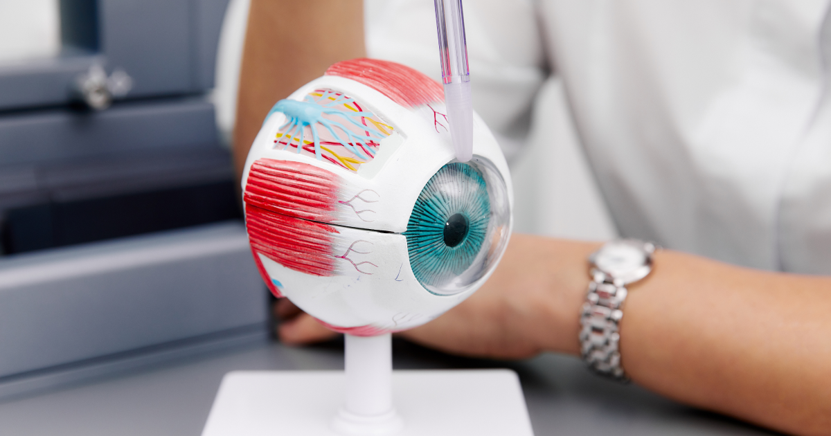
When people ask what is retina, they are usually trying to understand how a thin layer at the back of the eye helps create the detailed world we see. The retina is not just a screen which receives the image. It is live nerve tissue that which plays a role in the detection of light, the processing of color, contrast and motion and also which prepares visual inputs for the brain. Understanding the retina eye function can also make it easier to recognise why changes in vision sometimes require an eye examination.
At Vasan Eye Care, retinal health is an important part of comprehensive eye care because some retina problems may develop before they cause obvious symptoms. A planned retina check up which allows an ophthalmologist to study the macula, peripheral retina and optic nerve and determine if more in depth tests are required. This article explains what is retina in patient-friendly language while keeping the science accurate.
What Is Retina and Where Is It Located?
To answer what is retina, picture the eye as both an optical system and an extension of the nervous system. The retina is a thin out of which the light is put in the back of the eye. Light into the cornea, the pupil and the lens which in turn focus on this tissue. Specialized retinal cells then transform the light into electrical signals. These signals go through the optic nerve to the brain which in turn interprets them as the images we see.
An important part of understanding what is retina is recognising that it has organized layers of nerve cells. These cells do more than register light. They also identify edges, compare between dark and light areas, react to motion and put visual info into separate channels. This early processing is the base of retina eye function.
The macula is in the middle of the retina which in turn supports clear central vision. At the center of the macula is the fovea which has the best visual detail. The outer retina plays more of a role in peripheral vision and in low light. For this reason, different retina problems may affect central sight, side vision, night vision or visual clarity in different ways.
Main Structures Behind the Retina Eye Function
The answer to what is retina becomes easier when its main structures are understood. Each has a separate role, but all work together continuously.
| Retinal structure or cell | Main role | Simple explanation |
| Rod photoreceptors | Dim-light and peripheral vision | Rods are highly sensitive to light and help detect shapes and movement in darker surroundings. |
| Cone photoreceptors | Colour and fine detail | Cones support reading, face recognition and colour discrimination, especially near the macula. |
| Bipolar cells | Signal transfer and organisation | They carry information from photoreceptors towards ganglion cells through separate pathways. |
| Horizontal and amacrine cells | Contrast and motion refinement | They compare nearby signals and help highlight important visual differences. |
| Ganglion cells | Final retinal output | Their fibres form the optic nerve and carry processed signals towards the brain. |
| Macula and fovea | Detailed central vision | These areas support the sharp sight needed for reading, driving and recognising faces. |
This coordinated arrangement explains why the retina eye function involves much more than light detection. The retina processes, compares and organizes info before it leaves the eye. In the case of retina issues symptoms present in part which which areas or cells are affected.c
How Does the Retina Process Visual Information?
The simplest explanation of what is retina is that it works like a biological light sensor which also functions as an early image processor. To that which a camera does with its sensors the retina adds in that it is made up of live neurons which in turn adapt to changes in light and which constantly refine what the eye perceives.
Light Is Focused on the Retina
Visual input is a pre-retinal process. The cornea which is what does the light bending, the pupil which regulates the amount of light that gets in, and the lens which changes shape for focus. A small inverted image is projected on the retina but the brain reinterprets the info we send which in turn presents to us the world in its normal upright orientation.
Accurate focusing supports the retina eye function. Myopia, hyperopia or astigmatism may present with a blurred image which in fact may be of healthy retinal tissue. An exam can tell a focusing issue from a retina problems at the back of the eye. Accurate focus is what the retina requires.
Rods and Cones Convert Light Into Signals
Another key aspect of what is retina includes photoreceptors. Rods and cones which are the light sensitive elements. Upon light’s contact with these cells chemical and electrical changes which we term phototransduction take place. This is the process which turns light energy into messages that nerve cells then send.
Rods do well in low light and also play a key role in peripheral vision. Cones function better in bright light and also support color vision and fine detail. It is the combination of different cone types’ responses which allows the brain to tell between a large spectrum of colors. This conversion process is a core retina eye function. Damage to photoreceptors may lead to retina problems involving night vision, central detail, colour perception or the visual field.
Bipolar Cells Separate Light and Dark Information
Once the photoreceptors respond to light they pass that info on to bipolar cells. The retina doesn’t send out one whole picture to the brain. What it does is put out parallel paths of information. Some cells in this system react more so to an increase in light, while some react to a decrease in light. These are what we call ON and OFF pathways.
This is why what is retina cannot be answered only as “the layer that detects light.” It begins to put together the picture within the eye. An NCBI Bookshelf report notes that retinal circuits separate out info related to brightness, darkness, direction and rates of visual change before sending it to the brain. This organisation strengthens the retina eye function by helping us notice boundaries, letters, faces and moving objects.
Horizontal and Amacrine Cells Improve Contrast
To understand what is retina more deeply, it helps to know that neighbouring retinal cells communicate. Horizontal cells in fact compare input from close by photoreceptors, which in turn highlights the differences in light and dark areas. This process also plays a role in edge detection and contrast sensitivity.
Amacrine cells play a role in timing, movement and communication between bipolar and ganglion cells. This filtering supports the retina eye function and allows the optic nerve to carry refined information rather than a point-by-point copy of the scene.
Ganglion Cells Send Information to the Brain
Ganglion cells receive the processed output of the retinal network. Their long fibres join to form the optic nerve, which carries electrical impulses to visual centres in the brain.
When explaining what is retina, ganglion cells can be described as its final communication channel. What also is true is that different ganglion cells react to features like contrast, color and motion. Also the brain in this process puts together that which is reported by both eyes, memory and attention to produce what we experience as conscious sight. The retina eye function therefore depends on the retina, optic nerve and brain working together.
How Are Images Formed on the Retina?
The question what is retina is closely connected with image formation. Light which hits an object goes in through the cornea and the pupil and is brought to a focus by the lens. An inverted image forms on the retina. What the photoreceptors don’t do is send a picture to the brain; they take the pattern of light and turn it into a series of electrical and chemical signals.
The retina processes what it receives in terms of brightness, color, edges, contrast and movement. At that which is processed the ganglion cells report to the optic nerve, which in turn reports to the brain which creates the whole picture out of it. This pathway from focused light to perception is the practical meaning of the retina eye function.
Why Retina Problems Can Affect Vision Differently
Understanding what is retina also explains why retinal symptoms vary. Macular changes may cause blurring or distortion of central vision, at the same time peripheral changes may present as a shadow or loss of side vision. Photoreceptor issues may play with night vision or color perception. Edema, hemorrhage, tear or detach may bring on acute symptoms which in turn require prompt evaluation.
| Visual change | What it may suggest | Recommended response |
| Sudden flashes or a rapid increase in floaters | A retinal tear or vitreous traction may need to be ruled out | Seek urgent ophthalmic assessment. |
| A curtain, shadow or missing area in vision | Possible retinal detachment | Obtain emergency eye care without delay. |
| Wavy or distorted central vision | A condition involving the macula may be present | Arrange an eye examination promptly. |
| Gradual central blur | The cause may involve the retina, lens or focusing system | Book a comprehensive evaluation. |
| Reduced night or side vision | Retinal function or another visual pathway may be involved | Discuss the change during a retina check up. |
This table is to inform that it does not provide diagnosis of retinal conditions. Sudden vision loss, a new black curtain, rapid increase in flashes and floaters, or serious eye injury is a medical emergency. People with diabetes, high blood pressure, high myopia, previous eye surgery, a family history of retinal diseases or known retinal eye disease may need an examination schedule recommended by an ophthalmologist.
What Happens During a Retina Check Up?
A retina check up usually begins with a discussion of symptoms, medical history, medicines and past eye issues. Vision will be tested also we may use dilating drops which will allow the ophthalmologist to see a larger area at the back of the eye. We may use special lenses, slit lamp exam or retinal imaging which will be determined by what is needed clinically.
Optical coherence tomography, or OCT, can provide cross-sectional images of retinal layers. We also have fundus photography which documents the back of the eye. The exact retina check up depends on symptoms and suspected retina problems. It is also important to get a retina check up even when your vision seems fine which is when some issues may be quietly starting. At eye specialist hospital you can get a recommended exam schedule based on your age, symptoms and health risks.
When Should You Consult an Eye Specialist?
Knowing what is retina is useful, but knowing when to seek care is equally important. We may see new distortion, blurred central vision, difficulty with low light, sudden floaters, flashes, a shadow in what we see, or a sudden loss of vision which should not be ignored. While these signs do not always point to retina problems, they do require a visit for an in depth evaluation.
Patients which present to with retinal diseases should report for follow up as directed. People with diabetes, high blood pressure, previous retinal treatment, strong myopia or a family history of retinal eye disease should discuss monitoring with an ophthalmologist. Timely retina check up at an eye specialist hospital may support an earlier diagnosis and better eye treatment plan.
Protecting Retinal Health
Learning what is retina highlights the connection between general health and vision. We do well to follow as per doctor’s advice on blood sugar, blood pressure and cholesterol, to give up tobacco and to attend our scheduled exams which in turn support eye health. Home remedies can not take the place of a clinical retina check up when symptoms present. Retinal diseases may require watchful waiting, injections, laser treatment, surgery or management of an underlying condition. Care should be very personal at an eye specialist hospital.
Frequently Asked Questions
Rods and cones in the retina detect different aspects, retinal circuits then work out what is contrast, color and speed at the same time many neurons send those processed signals through the optic nerve to the brain.
In short the light which falls on our retina is transformed into bio-electrical messages and the brain interprets them into what we see including forms, colors, distance, and motion.
In bright conditions, sensitivity is generally highest around yellow-green light. In darkness, peak sensitivity shifts towards blue-green wavelengths.
The cornea and lens create an inverted image which is what hits the retina. That light pattern is converted by retinal cells into signals which the brain then interprets.
The retina in a nutshell is light sensitive nerve tissue at the back of the eye. It starts visual processing and sends info to the brain.
Usually a retina check up is not painful. May be a slight sting from the dilating drops which also may leave vision blurred or light sensitive for a few hours.
An ophthalmologist evaluates retinal diseases. Complex conditions may be managed by a retina specialist at an eye specialist hospital.
The answer to what is retina reveals one of the body’s most sophisticated sensory systems. It receives light, transforms it into electrical signals, sorts information into dedicated paths and puts forward a fine tuned report to the brain. Also in studying what is retina we see how it enables processes like reading, face recognition, color vision, motion detection and adjustment to varying levels of light.
Because retina problems can affect vision in different ways, persistent or sudden visual changes deserve professional evaluation. Understanding what is retina should lead to better health care decisions rather than self diagnosis. At Vasan Eye Care we provide full ophthalmic assessment and personal guidance for patients which require clarification of retina problems, symptoms or follow up care.
References
- NCBI Bookshelf, U.S. National Library of Medicine: How the Retina Works
https://www.ncbi.nlm.nih.gov/books/NBK610611/ - National Eye Institute, U.S. National Institutes of Health: Retinal Detachment
https://www.nei.nih.gov/eye-health-information/eye-conditions-and-diseases/retinal-detachment