
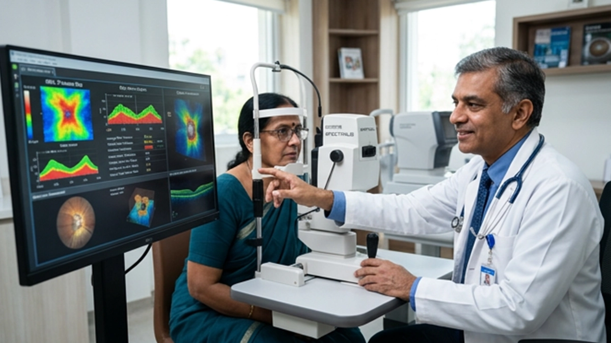
An OCT test for glaucoma which in detail produces images of the optic nerve and retinal layers. It is able to identify early signs of nerve tissue thin which may point to glaucoma before visual symptoms present. Also an OCT result by itself does not confirm glaucoma. Ophthalmologists will place the scan results in perspective with eye pressure, perform optic nerve exam, visual field test and also will factor in individual risk factors.
Glaucoma attacks the optic nerve and also may progressively dim peripheral vision. The National Eye Institute’s report on glaucoma notes that in the early stages of the disease many patients report no symptoms which is why we should all have full eye exams.
What Does an OCT Test for Glaucoma Examine?
OCT which stands for Reflective Light produces cross sectional images of structures at the back of the eye. In an OCT test for glaucoma the ophthalmologist pays close attention to the retinal nerve fibre layer, optic nerve head and macular ganglion cell region.
The RNFL which stands for Retinal Nerve Fiber Layer measures the tissue which surrounds the optic disc. This nerve fiber layer is made up of extensions of retinal ganglion cells which go on to form the optic nerve. In glaucoma this layer may become thinner.
An optic nerve oct can also does assessment of the optic disc structure and areas of neuroretinal rim loss. At Vasan Eye Care we have a glaucoma info page which patients may find of use.
| OCT measurement | What it examines | Why it matters in glaucoma |
| RNFL thickness | Nerve fibre layer around the optic disc | Localised or progressive thinning may indicate glaucomatous damage |
| Ganglion-cell analysis | Retinal ganglion-cell layers near the macula | May reveal structural nerve-cell loss |
| Optic nerve head | Disc, rim and cup characteristics | Helps assess structural changes associated with glaucoma |
| Serial comparison | Changes between scans over time | Helps determine whether structural damage is progressing |
How Is the OCT Glaucoma Test Performed?
The OCT glaucoma test is a non-invasive procedure which usually takes a few minutes. The patient places their chin and forehead against the machine which is to look at a fixed target. The device scans each eye without touching it.
Pupil dilation at times will improve image clarity. If there is a cataract, dry eye, blinking or poor fixation issues which interfere with the scan that is when we see a drop in image quality. At Vasan Eye Care we have detailed out the wide range of uses and the process of OCT in our scan guide. An OCT test for glaucoma is very much a part of a comprehensive glaucoma eye test and not a separate investigation.
What Do RNFL and OCT Results Mean?
A retinal nerve fiber layer test report usually compares what was measured with a reference database. Also many OCT systems present color coded classifications. The colors may aid in interpretation but they do not in and of themselves provide a diagnosis.
Research reports that RNFL assessment is a valuable tool. In 2026 a study which did a systematic review and network meta analysis of 47 studies with 8,177 participants and 12,723 eyes which came up with that average and inferior RNFL measurements had the highest diagnostic accuracy of reported at 0.77.
| Finding | Possible interpretation | Important limitation |
| RNFL within expected range | No obvious structural loss detected | Does not completely exclude early glaucoma |
| Localised RNFL thinning | May raise suspicion of glaucoma | Anatomy and scan artefacts can mimic thinning |
| Ganglion-cell thinning | May support structural nerve damage | Must match other clinical findings |
| Progressive thinning | May suggest disease progression | Reliable comparison requires good-quality serial scans |
The ophthalmologist looks at trends instead of a single figure. In some cases of glaucoma OCT scan we may see abnormal results which are a result of high myopia, anatomical variation, retinal disease or imaging artifacts. Age also is a factor in RNFL thickness.
For this reason, an oct test for glaucoma should always be interpreted clinically.
Why OCT Is Not Enough to Diagnose Glaucoma
Glaucoma diagnosis is a process which includes structural and functional assessment. OCT is used for tissue structure assessment, on the other hand visual field testing looks at how glaucoma affects vision. In some cases eye pressure is a factor although it is true that glaucoma can present without very high intraocular pressure.
Tonometry is that which measures intraocular pressure and a dilated eye exam is performed to evaluate the optic nerve directly. Thus a full eye exam may include OCT, tonometry, optic nerve study and visual field testing. People with known risk factors for glaucoma may require more frequent monitoring.
Can an OCT Test for Glaucoma Detect Progression?
Yes. Repetitive scans are a tool which ophthalmologists use to identify progressive RNFL or ganglion cell thinning. We see that serial comparison is a better approach than looking at a single OCT test for glaucoma. Progression software which looks at data from various visits is used. Also the ophthalmologist has to check image quality, segmentation errors and that the change follows a glaucoma pattern.
Patients also should be made aware of the first signs of glaucoma. Sudden severe eye pain, blurred vision, a red eye, nausea or halos around lights require prompt ophthalmic assessment as acute angle closure glaucoma is a medical emergency.
Eye Care at Vasan Eye Care
Vasan Eye Care offers assessment of glaucoma and other optic-nerve conditions. As per clinical evaluation which may include a wide range of tests, an ophthalmologist may recommend an OCT test for glaucoma along with measurement of pressure, visual field testing and dilated exam.
For best results bring in previous OCT reports to your follow up visits as we may be able to identify what has changed over time. We have a directory of eye hospitals which will help you to find a convenient center.
Related Glaucoma Tests and Eye Conditions
An OCT scan helps eye care professionals examine the optic nerve and retinal nerve fibre layer, supporting the assessment and monitoring of glaucoma. To understand the condition better, explore the common glaucoma causes and learn about optic nerve swelling symptoms, which may indicate a separate optic nerve condition. You can also discover the difference between glaucoma and cataract to understand how these two eye diseases affect vision differently. Read about diplopia to learn more about double vision, its possible causes, and evaluation. If you experience discomfort or causes of eye pressure pain, an eye examination can help identify the underlying reason. For more information about glaucoma assessment, explore the glaucoma eye test and consult an eye care professional about the tests appropriate for your symptoms and risk factors.
Frequently Asked Questions
1. Can an OCT test for glaucoma confirm the diagnosis?
No. An OCT scan for glaucoma gives out valuable structural info, but OCT by itself is not enough to diagnose or rule out glaucoma. Ophthalmologists put the scan with the optic nerve exam, intraocular pressure, visual field results, medical history and other related factors.
2. What does RNFL thinning mean on an OCT report?
RNFL atrophy which means the retinal nerve fibre layer is thinner in a given area. Glaucoma causes this to present but also normal anatomy, myopia, retinal diseases and scan artifacts may play a role in results.
3. How often should an OCT glaucoma scan be repeated?
There is not a uniform schedule that fits all. What we see again and again is that which tests to repeat is based on the degree of glaucoma, risk factors, past results and also the presence of disease progression. An ophthalmologist will determine the monitoring interval after looking at the whole picture.
4. Is the OCT test for glaucoma painful?
No. An OCT test for glaucoma usually does not contact the eye and uses light which is as opposed to radiation. The scan itself is very quick. At times dilating drops may be required which in turn may temporarily cause close vision to blur or light sensitivity to increase.
Persistent and progressive changes in vision should be paid attention to. An ophthalmologist is able to look at the optic nerve, analyze OCT results and to determine if it is a case of monitoring or glaucoma treatment. Patients may go to a Vasan Eye Care center for a full evaluation.
References
- National Eye Institute — Glaucoma
https://www.nei.nih.gov/eye-health-information/eye-conditions-and-diseases/glaucoma - Early Diagnosis of Glaucoma By Optical Coherence Tomography: A Systematic Review and Network Meta-analysis
https://pubmed.ncbi.nlm.nih.gov/41815442/