
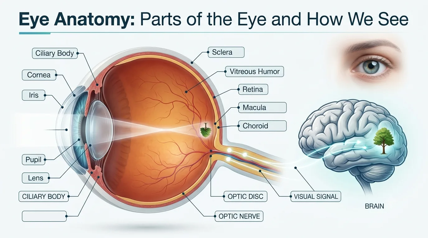
Our senses are what we use to experience the world which includes recognizing faces, reading, working, driving, and enjoying day to day events. Though vision may seem like a simple function, it is a very complex process which has many elements that work in perfect unison. By learning about human eye anatomy you may better understand how your vision works and also the importance of having regular eye exams for a lifetime of good eye health.
Whether you have questions about the parts of the eye and their functions or are interested in the function and structure of the eye, this guide goes over the anatomy of the human eye in simple, easy to understand terms.
What Is Human Eye Anatomy?
Human eye anatomy structures which present the eye’s parts that work as a team to capture light, convert it to electrical signals, and send those signals to the brain which in turn interprets them as images
The eye is a camera. A camera takes photos, the eye instead which is a living entity, it constantly changes focus, brightness, color, and depth perception. Each component has a different role and in the event of even minimal damage to that structure vision is affected.
Knowledge of the function and structure of eye also has a role in which patients recognize that different eye conditions may require different eye treatments and that early diagnosis by an experienced eye doctor is key.
Parts of the Eye and Their Functions
The human eye consists of several interconnected structures that contribute to clear vision.
| Part of the Eye | Primary Function |
| Cornea | Bends incoming light and provides most of the eye’s focusing power. |
| Iris | Controls the amount of light entering the eye by adjusting pupil size. |
| Pupil | Opening that allows light to enter the eye. |
| Lens | Fine-tunes focus for near and distant objects. |
| Retina | Converts light into electrical signals. |
| Optic Nerve | Carries visual information from the retina to the brain. |
| Vitreous Humour | Maintains the eye’s shape and supports the retina. |
Cornea
The cornea which is the front of the eye is transparent. It plays the role of the first lens which is to refract the incoming light. Also because it does not have blood vessels it is clear which in turn allows for efficient passage of light.
Injuries, infection, or disease of the cornea may cause blurred vision, pain, or light sensitivity which in turn may require special eye treatments.
Iris
The iris which is the colored part of the eye. In it are very small muscles which control the size of the pupil in response to light.
In full sunlight the pupil reduces in size to let in less light. In poor lighting which is dim the pupil dilates to let in more light.
Pupil
In bright sunlight the pupil reduces in size to let in less light. In dim lighting the pupil dilates to let in more light.
The pupil’s size is in a constant state of change for the purpose of producing clear and comfortable vision.
Lens
In the rear of the iris the natural lens changes its shape in order to focus light onto the retina. This process which we term accommodation also allows us to switch focus between near and far objects.
As we age our lenses lose flexibility which in turn causes presbyopia, a very common vision issue at an older age which affects near vision.
Retina
The retina which is a very thin layer of nerve tissue that lines the back of the eye. It also includes special photoreceptor cells which we know as rods and cones
Rods play a role in low light which we see, while cones are responsible for color vision and sharp central vision.
According to the American Academy of Ophthalmology the retina transforms light into electrical signals which then travel through the optic nerve to the brain for image processing.
Macula
At the base of the retina sits the macula which in turn is responsible for central detailed vision. We use it for activities like reading, recognizing faces, and driving which all benefit from a healthy macula.
Optic Nerve
The optic nerve is the communications path between the eye and the brain. It transmits visual information from the retina.
Damage from glaucoma which affects the optic nerve may cause permanent vision loss without treatment.
Vitreous Humour
The vitreous humor is a clear gel which fills the space between the lens and the retina. It plays a role in maintaining the eye’s shape and supporting internal structures.
As we age the vitreous changes which in turn may cause floaters or more rarely retinal tears.
How Do We See?
Understanding of human eye anatomy is achieved when you know how vision works.
Light first hits the cornea which in turn passes it to the pupil. The iris which is responsible for that which we see as the color of the eye also changes the pupil size in response to light levels. The lens then does a fine job of tuning light focus, sending it precisely to the retina.
In the retina we have rods and cones which convert light into electrical signals. These signals go through the optic nerve to the brain’s visual centers at which point they are interpreted into the images we see.
The U.S. National Library of Medicine (MedlinePlus) reports that what we see is a result of this intricate play between the eye and brain which in turn allows us to see color, depth, movement, and detail.
Understanding the Function and Structure of Eye
The function and structure of eye are related. Each element plays a role in producing clear, comfortable vision.
| Eye Structure | Everyday Activity Supported |
| Cornea | Reading, driving, watching television |
| Iris and Pupil | Adjusting to bright sunlight and dark rooms |
| Lens | Reading books and viewing distant objects |
| Retina | Colour recognition and detailed vision |
| Macula | Face recognition and fine tasks |
| Optic Nerve | Sending visual information to the brain |
| Vitreous Humour | Maintaining eye shape and retinal support |
Common Conditions That Affect Eye Anatomy
Several eye conditions which do to their nature interfere with function and structure of eye.
Cataracts which is when the natural lens becomes opaque cause blurred vision.
Glaucoma which is a result of damage to the optic nerve mostly from increase in eye pressure.
Diabetic retinopathy we see as a result of damage to blood vessels in the retina which in turn is a result of diabetes.
Age related macular degeneration which mainly affects central vision as a result of macula damage.
Corneal infections or injuries which in turn may cause the cornea to not focus light properly.
Each of these conditions affect different parts of the eye and their functions which is why accurate diagnosis is key.
Symptoms That Should Never Be Ignored
Although many eye issues present slowly, some symptoms require a timely evaluation by a health professional with experience in eye doctor.
Persistent blurry vision, sudden loss of vision, flashes of light, new floaters, eye pain, redness, double vision, increased sensitivity to light, night time vision impairment should be paid attention to .
Early detection tends to improve results of treatment and also preserve vision.
How Eye Problems Are Diagnosed
A full eye exam assesses the total health of the eye and its components.
Based on your symptoms, an eye doctor does a visual acuity test, slit lamp exam, retinal evaluation which may require pupil dilation, intraocular pressure measurement, Optical Coherence Tomography (OCT), or retinal imaging.
In an eye specialist hospital we have at our disposal modern diagnostic technologies which detect many eye conditions before vision is lost.
Treatment Options
Treatment is based on what part of the eye is affected.
For minor infections we may use medicated eye drops and for refractive errors we correct with glasses or contacts. Cataracts usually require surgery as soon as vision is very impaired. In case of glaucoma we manage by using medications, laser, or a surgical procedure to lower eye pressure. Retinal issues may require injections, laser treatment, or vitreoretinal surgery. Of all of these the appropriate eye treatments are determined by in depth examination from a trained ophthalmologist.
Can Eye Problems Be Prevented?
Although there is no way to prevent all eye diseases, adopting healthy habits greatly reduces your risk of vision problems.
Regular routine eye exams, also at age 40 and beyond in the case of diabetics and those with a history of eye disease, which help to identify issues early.
A balanced diet that includes lots of leafy greens, fruits, and omega-3’s is good for retinal health. Also wear protective eye wear while playing sports or at work which is dangerous to the eyes, manage diabetes and blood pressure, do not smoke, which goes for second hand as well, and limit screen time, and follow the 20-20-20 rule which is looking away from your screen every 20 minutes for 20 seconds at something 20 feet away.
Routine trips to an eye specialist hospital is a very effective method of protecting long term eye health.
When Should You Consult an Eye Specialist?
Consult an eye doctor which deals with the eyes if you notice changes in vision, persistent eye discomfort, frequent headaches related to visual tasks, or any sudden visual symptoms.
Children should have their eyes checked at regular intervals as early detection of vision issues supports healthy visual and cognitive development.
Timely evaluation also allows for the right eye treatments to be given before issues worsen.
Conclusion
Understanding human eye anatomy which in turn allows you to see how each component plays a role in overall vision. As each element of the eye has a specific job, it is important to have regular eye exams which in turn help in the early detection of issues and in the protection of your eyes over a lifetime
If you see changes in your vision or have issues related to your eye health, consult a reputable ophthalmologist at our trusted eye specialist hospital. At Vasan Eye Care we have a team of professionals which provide in-depth eye exams and tailored eye treatments to help you maintain healthy vision at every stage of life.
Frequently Ask Questions
The eye is made up of the cornea, iris, pupil, lens, retina, optic nerve, and vitreous humour. These structures work together to allow light in, focus it properly, and send visual info to the brain.
The eye which receives light, forms images, controls what light enters in, determines color, enables night vision, supports depth perception, has peripheral vision, detects motion, transmits visual info to the brain, and also plays a role in balance through visual orientation.
The iris controls the amount of light that enters the eye by means of the pupil’s size. It automatically changes in response to which is it very bright or dim out which helps maintain vision clear and continuous.
The iris controls the amount of light that enters the eye by means of the pupil’s size. It automatically changes in response to which is it very bright or dim out which helps maintain vision clear and continuous.
The base of the eye includes the cornea, iris, pupil, lens, retina, macula, optic nerve, sclera, conjunctiva, and vitreous humour. These structures work together to produce clear and detailed vision.