
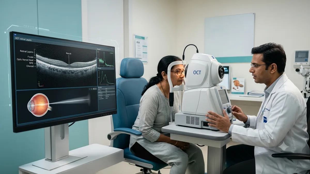
Healthy vision also depends on the early identification of eye diseases which often develop without any noticeable symptoms at first. In the field of modern ophthalmology we see very advanced imaging technologies among which the OCT eye test, stands out, which allows us to look at the retina in great detail without even touching the eye doctor
Whether you suffer from diabetes, glaucoma, age related macular degeneration, or unidentified vision changes, an OCT eye exam is a tool which may identify issues at early stages of development, often before permanent vision loss has a chance to take place. This non-invasive scan has also become a key element in the diagnosis and management of a number of retinal and optic nerve conditions.
In this guide we will go over what is OCT, how the optical coherence tomography test works, who is a candidate for it, and what to expect during the procedure.
What Is an OCT Eye Test?
An OCT eye test which stands for Optical Coherence Tomography is a pain free imaging scan that uses light waves to produce very detailed cross sectional images of the retina, the light sensitive tissue at the back of the eye.
Think of it as a modern twist of the ultrasound, what we have here is a technology that uses reflected light which produces high resolution images of various retinal layers. This in turn allows an eye doctor to identify fine structural changes which may go unnoticed in a regular eye test.
According to the American Academy of Ophthalmology (AAO) Optical Coherence Tomography is a key tool in diagnosis and management of retinal diseases, glaucoma and optic nerve disorders.
What Does Optical Coherence Tomography Mean?
If you were curious as to what OCT is, it stands for Optical Coherence Tomography.
What OCT does is that it gives us in depth pictures of the retina, optic nerve, and macula through the use of safe infrared light. Different from X-rays or CT scans, OCT does not use radiation at all.
Also the scan allows ophthalmologists to measure retinal thickness, identify swelling, see into the fluid accumulation which may be present, and track a disease’s progression over time.
How Does an OCT Eye Exam Work?
During the OCT eye exam you will sit at the testing machine which will have a support for your chin. You will be asked to look at a target while the scan takes in multiple images within seconds.
The machine does not touch your eye at all, and the scan is pain free. In some cases we may use dilation of your pupils to get better retinal images, though dilation isn’t always required.
The retinal layers are imaged which in turn allows the eye doctor to see very early stages of disease.
OCT Eye Test at a Glance
| Feature | Details |
| Test Type | Non-invasive retinal imaging |
| Technology Used | Light waves (Optical Coherence Tomography) |
| Duration | Usually 5–10 minutes |
| Pain | Completely painless |
| Radiation | None |
| Contact with Eye | No |
| Immediate Results | Images available immediately for review |
Why Is an OCT Eye Test Performed?
An OCT eye test is recommended for in depth evaluation of the retina or optic nerve. It allows specialists to see disease before symptoms of vision loss present.
Your eye doctor may recommend an OCT eye exam if you have:
- Blurred or distorted vision
- Diabetes
- Glaucoma
- Age-related macular degeneration
- Sudden loss of vision
- Retinal swelling
- Macular hole
- Optic nerve disorders
- High myopia with retinal concerns
- Unexplained visual symptoms
It is also a common practice to use it for follow up of certain eye care procedures in which we evaluate the eye treatments results.
What Diseases Can an OCT Eye Test Detect?
optical coherence tomography test is a very useful tool in ophthalmology which is able to identify many eye diseases in the early stage.
These include:
Age-Related Macular Degeneration (AMD)
OCT is able to detect fluid leakage, retinal swelling, and abnormal blood vessel growth related to macular degeneration which in turn will occur before severe vision loss.
Diabetic Macular Edema
People with diabetes can develop macular swelling. OCT which measures retinal thickness very well also allows for early diagnosis and treatment planning.
Glaucoma
OCT also measures the retinal nerve fiber layer and optic nerve health which in turn enables the early detection of glaucoma related nerve damage.
Macular Hole
Tiny details of the macula are brought out by OCT which in turn helps in the assessment of the severity and of the appropriate management.
Epiretinal Membrane
Scar tissue that develops on the retina will cause distortion in vision. OCT identifies these changes and measures the progression.
Retinal Detachment
Certain retinas’ pathologies including detachments may be studied with OCT also in association with the clinical evaluation.
The National Eye Institute reports that early diagnosis of retinal diseases is key to timely management and preservation of vision.
What Is Optical Coherence Tomography for the Macula?
The macula is that which is at the center of the retina which we use for what is fine detail in our vision for things like reading, writing, and to see faces.
An optical coherence tomography test focused on the macula evaluates:
- Retinal thickness
- Fluid accumulation
- Macular swelling
- Macular degeneration
- Macular holes
- Epiretinal membranes
This specialized scan is also very useful for patients with diabetes or age related macular degeneration
Who Should Consider an OCT Eye Exam?
Although OCT is not needed at every eye test, it can be particularly helpful for people who:
- Have diabetes
- Are over 40 years of age
- Have glaucoma or a family history of glaucoma
- Experience unexplained vision changes
- Have retinal disease
- Require monitoring after retinal eye treatments
- Have high-risk eye conditions identified during routine examination
Your eye doctor will determine which scans are necessary based on your symptoms and clinical reports.
Common Conditions Evaluated Using OCT
| Eye Condition | How OCT Helps |
| Diabetic Macular Edema | Detects retinal swelling and fluid |
| Glaucoma | Measures optic nerve and nerve fibre thickness |
| Macular Degeneration | Identifies abnormal retinal changes and fluid |
| Macular Hole | Visualises hole size and depth |
| Epiretinal Membrane | Detects retinal surface distortion |
| Retinal Vein Occlusion | Evaluates retinal swelling and treatment response |
Is an OCT Eye Test Safe?
Yes. An OCT eye test is very safe.
We use light in this test which also means it is fine to have it done many times over a few months or even years. There is no injection, no touching of the eye or use of surgery instruments.
If we do use dilating drops for the pupil, you may notice some blur and light sensitivity for a few hours.
Preparing for an OCT Eye Exam
Most people do not have to do any special preparation for an OCT eye exam.
If your pupils will be dilated you may prefer to have someone else drive you home which may be the case that you are uncomfortable at the time of the procedure. Also bring your current glasses or contact info as that will be useful at the eye hospital facility.
Understanding Your OCT Results
After performance of the optical coherence tomography test, the images are evaluated by your eye doctor.
The scan may reveal:
- Normal retinal layers
- Areas of retinal swelling
- Fluid beneath the retina
- Nerve fibre thinning
- Macular abnormalities
- Structural changes requiring monitoring
OCT results are evaluated in conjunction with your symptoms, vision exam, and other eye test results before we determine the treatment.
Can OCT Replace a Routine Eye Examination?
No.
While the OCT eye test which does very in depth retinal imaging is a great tool we use in addition to the full scale eye exam. In a full eye test we do vision testing, check eye pressure, do a slit lamp exam, and also evaluate the retina.
These assessments, taken together, support an accurate diagnosis and a personalised treatment plan.
When Should You Visit an Eye Hospital?
Seek prompt evaluation at a trusted eye hospital if you notice:
- Sudden blurred vision
- Distorted central vision
- Flashes of light
- New floaters
- Loss of side vision
- Difficulty reading despite updated glasses
- Vision changes associated with diabetes
Early diagnosis often allows more effective management and helps protect long-term vision.
Conclusion
Many of what we term serious eye conditions present with no apparent symptoms in their early stages. We are able to get very detailed images of the retina and the optic nerve which in turn allows for earlier diagnosis, better monitoring, and more informed treatment with the help of an OCT eye test. In conjunction with a full eye test this advanced imaging technology plays a role in preserving long term vision.
If you are a diabetes patient, have glaucoma, age related macular degeneration, or are seeing changes in your vision, we recommend that you see a qualified eye doctor at a trusted eye hospital like Vasan Eye Care for a tailored evaluation and advice on best eye treatments solutions for your health issues.
Frequently Ask Questions
An OCT eye test is very safe with no report of major side effects as it uses harmless light. In the event that pupil dilation is done temporary blurred vision and sensitivity to light may present for a few hours.
An optical coherence tomography test for the macula which produces in depth images of the central retina and which in turn is used to diagnose macular degeneration, diabetic macular edema, macular holes and other diseases which affect central vision.
The price of an OCT eye test is variable by the eye hospital facility, city and also what other tests may be necessary. We recommend you check in with your health care provider for exact prices at the time of your visit.
No. An OCT eye exam does not cause any pain and is non-invasive. The scanner does not come into contact with the eye, and the imaging process is usually for a few minutes.
An OCT eye test is used to detect glaucoma, diabetic macular edema, age related macular degeneration, macular holes, epiretinal membranes, retinal swelling and also many other retinal and optic nerve disorders.
The aim of the OCT eye test is to present in great detail the retina and optic nerve which in turn allows for early detection, diagnosis, monitoring and treatment of many eye diseases.
OCT is a term used for Optical Coherence Tomography which is an advanced imaging technology that produces in depth cross sectional images of the retina using light waves.
Yes. When an eye doctor recommends it, an OCT eye exam provides info which may detect eye disease at an earlier stage than what we see in a routine exam which in turn supports early diagnosis and proper management.
References link
- https://www.aao.org/eye-health/treatments/what-is-optical-coherence-tomography
- https://www.nei.nih.gov/learn-about-eye-health
- https://medlineplus.gov/lab-tests/optical-coherence-tomography-oct/
- https://www.nhs.uk/conditions/macular-degeneration/
- https://www.cdc.gov/diabetes/diabetes-complications/eye-health.html