
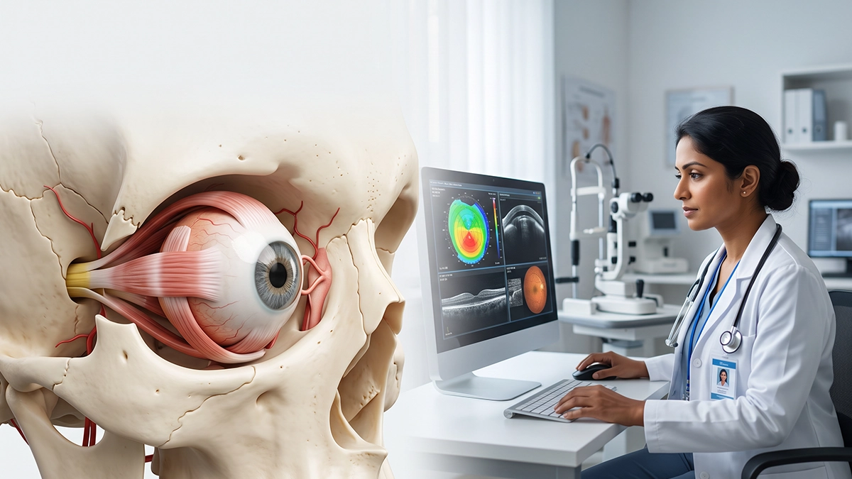
The eye orbit is the bony structure which contains and protects the eye ball. In it also are included muscles, nerves, blood vessels, fat, and tear related structures which play a role in normal eye function. Knowledge of the eye orbit is important in discussion of facial injuries, swelling, double vision, or changes in eye position. The adjacent eye socket bone structure which forms the eye socket plays a role as a protective framework for these delicate tissues.
What Is the Eye Orbit?
The eye orbit which is what we also call the eye socket is a cone shaped cavity in the head within the skull which houses the eye and the related tissues. The NCBI Bookshelf reports that in the orbit you will find the eye globe, the optic nerve, extraocular muscles, vessels, the lacrimal gland, fat and connective tissue. Also present are foramen which allow nerves and blood vessels to pass between the eye, face and brain.
The socket is made up of seven different bones instead of what we may think of as a single eye socket bone.
Bones That Form the Eye Orbit
The eye orbit has a roof, base, inner wall, and outer wall. Together which walls form a strong protective socket.
| Bone | Main contribution |
| Frontal and sphenoid | Form much of the roof and back |
| Maxilla, zygomatic and palatine | Contribute mainly to the floor |
| Ethmoid and lacrimal | Help form the medial wall |
| Sphenoid and zygomatic | Support the lateral wall |
A fracture of the eye socket bone may occur in a fall, from a car accident, a sports injury, or a direct hit to the face. The socket is near the sinuses, nerves, and eye muscles which means that some fractures may in turn affect movement or vision.
Muscles and Nerves Inside the Eye Orbit
Extraocular muscles present in the eye orbit move the eyes up, down, in and out and also in rotation. Also in the orbital socket we find cranial nerves which control sight, eye movement, eyelid elevation and sensation.
| Structure | Main role |
| Optic nerve (CN II) | Carries visual information to the brain |
| Cranial nerves III, IV and VI | Control most eye movements |
| Ophthalmic division of CN V | Provides sensation around the eye |
| Extraocular muscles | Move and align the eyeball |
| Lacrimal gland and orbital fat | Support tears and cushion the eye |
In that which is a small space many structures share, disease within the eye orbit may cause double vision, restricted eye movement, numbness, eyelid changes, or reduced vision.
Common Disorders Affecting the Eye Orbit
Conditions of the eye orbit which we see include orbital fractures, infections, inflammation, thyroid eye disease, cysts, vascular anomalies, and tumors. A broken eye socket bone may disrupt nearby muscles. Infection from the sinuses may get into the orbital socket, also inflammation may cause swelling of the eye to push it forward.
The eye orbit which also includes the optic nerve means that a quick increase in pressure is a risk to sight. Sudden vision loss, severe pain, fever, new double vision, or an eye that is protruding requires prompt medical attention.
How Are Eye Orbit Problems Diagnosed?
An eye specialist may test vision, pupils, eye movement, eyelid function, for swelling and note the position of each eye. If there is a problem with the eye orbit we may order a CT or an MRI. CT is best for assessment of a bone injury in the eye socket bone and the MRI gives us detailed images of the nerves and soft tissues.
Treatment is based on what is causing the issue. In the case of minor injuries you may only require to be watched, but we may also use medicine, perform a procedure or refer you to a specialist for issues like infection, serious fractures, inflammation, nerve pressure, or a mass.
Eye Care at Vasan Eye Care
Pain, swelling, double vision, abnormal eye movement, or vision changes as a result of a facial injury should be presented to an eye hospital. At Vasan Eye Care an eye specialist can look at whether adjacent orbital tissues are affected and will order appropriate tests based on what is seen. We stress the importance of early eye care when symptoms present suddenly or are getting worse.
Conclusion
The eye is protected by the eye orbit which also supports normal movement, sensation, blood supply, and nerve function. The orbital socket works with nearby bones and soft tissues to create a compact yet very organized space. Injuries and swelling to the eye area require prompt medical evaluation.
Frequently Asked Questions
1. Is the eye orbit the same as the eye socket?
Yes. The eye socket is a term that is used to refer to what is also known as the eye orbit.
2. How many bones form the eye orbit?
The eye orbit which also includes is made up of the frontal, sphenoid, zygomatic, maxilla, ethmoid, lacrimal, and palatine bones.
3. Can an eye socket fracture affect vision?
Yes. A broken bone may also injure nearby muscles, nerves or tissues and in some cases may cause double vision or other visual signs.
4. What is inside the orbital socket?
The orbital socket includes the eyeball, optic nerve, eye muscles, vessels, lacrimal gland, fat, and supportive tissues.
5. When should I seek urgent eye care?
Seek immediate eye care for acute vision loss, severe pain, fever, quick onset of swelling, an eye that protrudes, or the onset of double vision.
References
- NCBI Bookshelf – Anatomy, Head and Neck, Orbit Bones https://www.ncbi.nlm.nih.gov/books/NBK531490/
- NCBI Bookshelf – Anatomy, Head and Neck, Orbit https://www.ncbi.nlm.nih.gov/books/NBK539843/