
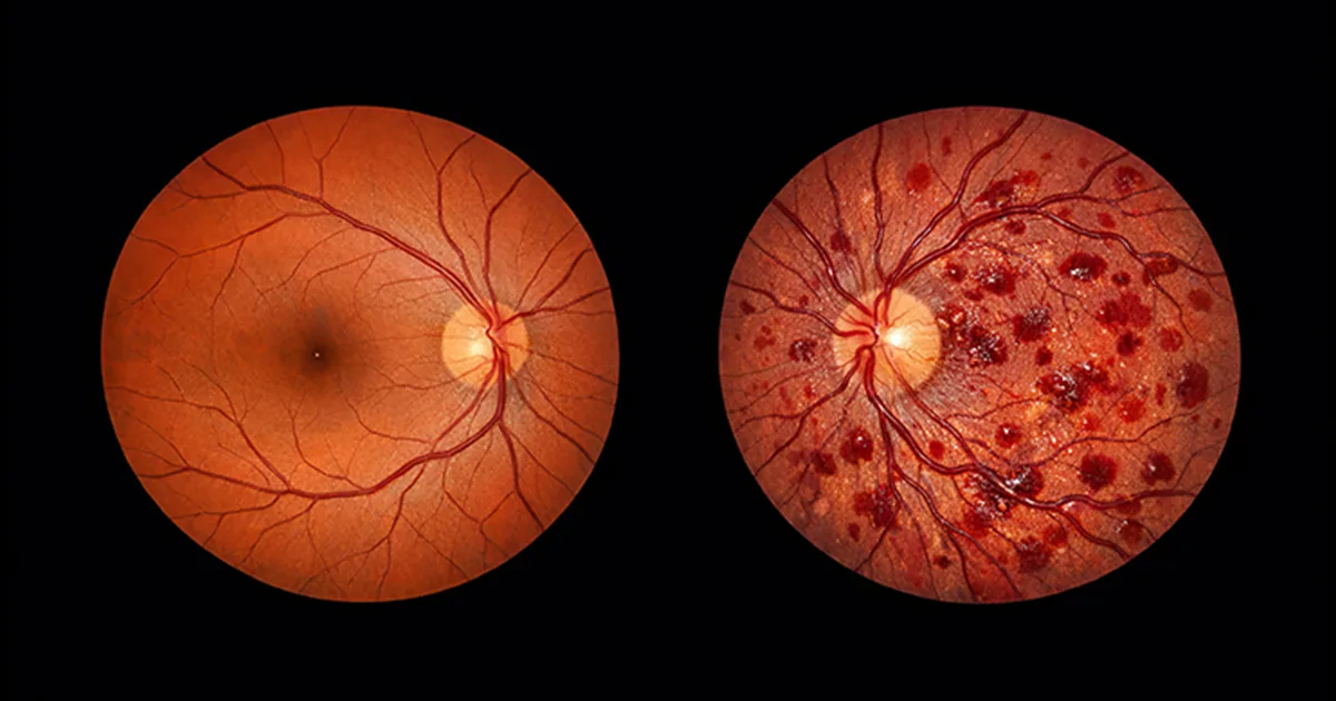
During a dilated eye examination, your retina specialist might point to a handful of tiny, dark red spots on the retina. The formal name is dot and blot hemorrhages. They do not usually cause dramatic vision loss on their own, but they are important signs. Most often, they signal diabetes or hypertension quietly affecting the small vessels at the back of the eye.
This guide walks you through what dot and blot hemorrhages are, what causes them, how they are diagnosed, and what treatment options exist.
What Are Dot and Blot Hemorrhages?
Dot and blot hemorrhages are small, rounded haemorrhages in the deeper layers of the retina (inner nuclear and outer plexiform layers). They appear dark red or reddish-brown on fundus examination.
- Dot hemorrhages: smaller, sharper, and more discrete
- Blot hemorrhages: slightly larger, less defined
They are different from flame-shaped haemorrhages, which sit in the surface nerve fibre layer and are often linked with high blood pressure.
What Are the Four Types of Retinal Hemorrhages?
A practical summary.
1. Intraretinal (dot and blot) hemorrhages
Deeper retinal layers; common in diabetic retinopathy.
2. Flame-shaped hemorrhages
Superficial nerve fibre layer; common in hypertensive retinopathy.
3. Preretinal hemorrhages
In front of the retina, between retina and vitreous.
4. Vitreous hemorrhages
Bleeding into the vitreous gel; causes sudden floaters and blurred vision.
Some systems also include subretinal hemorrhages (under the retina) and choroidal hemorrhages (in the choroid layer).
What Causes Dot and Blot Hemorrhages?
1. Diabetes (diabetic retinopathy)
The commonest cause globally. Chronic high blood sugar damages the small retinal vessels.
2. Hypertension
Raised blood pressure damages retinal vessels and can cause a mix of hemorrhage types.
3. Retinal vein occlusion
Blockage of a retinal vein causes widespread dot, blot, and flame hemorrhages in the affected area.
4. Retinal artery occlusion
Sudden blockage of a retinal artery can also cause some hemorrhages alongside ischaemic changes.
5. Anaemia
Severe anaemia can cause dot and blot hemorrhages.
6. Blood disorders
Thrombocytopenia, leukaemia, and other haematological conditions can cause retinal bleeding.
7. Valsalva retinopathy
Forceful straining, coughing, or lifting can rupture small vessels.
8. Trauma
Direct eye injury or sudden deceleration.
9. Shaken baby syndrome
In infants; an important paediatric emergency.
10. Coagulation disorders
Blood-thinner medicines in excess or coagulation factor deficiencies.
11. High altitude
High-altitude retinopathy can produce dot and blot hemorrhages.
12. Infections
Certain viral infections can cause retinal bleeding in select cases.
Symptoms of Dot and Blot Hemorrhages
- Often asymptomatic
- Mild blurred vision if the macula is affected
- Occasional small blind spots
- New floaters if nearby vessels bleed into the vitreous
- Gradual loss of clarity over time in chronic cases
- Sudden change if a large hemorrhage occurs
Many patients discover them during a routine diabetic eye check rather than from symptoms.
How Are Dot and Blot Hemorrhages Diagnosed?
A thorough evaluation at an eye specialist hospital includes:
- Visual acuity
- Eye pressure
- Slit-lamp examination
- Dilated fundus examination
- Fundus photography
- Optical coherence tomography (OCT) of the macula
- Fluorescein angiography to map blood flow
- Blood tests: HbA1c, blood pressure, complete blood count, coagulation, lipids
- Systemic medical review
Clinical grading of diabetic retinopathy helps decide follow-up and treatment intervals.
How Serious Is a Macular Hemorrhage?
A macular hemorrhage is bleeding in the central, detail-vision area of the retina. It can cause significant central vision loss depending on size, depth, and duration. Common causes include:
- Diabetic macular oedema
- Neovascular age-related macular degeneration
- Macroaneurysm rupture
- Vein occlusion
- Trauma
Macular hemorrhages always need prompt specialist review and targeted treatment, which may include injections, laser, or surgery.
How Are Dot and Blot Hemorrhages Treated?
Treatment targets the underlying cause and any complications.
1. Diabetic retinopathy
- Tight blood sugar control
- Blood pressure and cholesterol management
- Anti-VEGF injections for macular oedema
- Laser photocoagulation in proliferative disease
- Vitrectomy for complicated bleeding
- Structured retinal disease treatment pathways tailor the approach to each patient
2. Hypertensive retinopathy
- Strict blood pressure control
- Monitoring retinal changes
- Managing cardiovascular risk
3. Retinal vein occlusion
- Anti-VEGF injections for macular oedema
- Laser in selected cases
- Treatment of systemic risk factors
4. Anaemia and blood disorders
- Treating the systemic condition
- Monitoring retina until resolution
5. Valsalva or traumatic hemorrhages
- Observation in many cases
- YAG laser or vitrectomy for persistent preretinal or vitreous bleeds
6. High-altitude retinopathy
- Descent and supportive care
Supportive eye treatments including lubricating drops and careful examination schedules accompany specific therapy. Patients with broader retinal conditions benefit from integrated retinal diseases care pathways.
Follow-up Intervals
- Mild NPDR with few hemorrhages: 6-12 months
- Moderate NPDR: 3-6 months
- Severe NPDR: 2-4 months, often with treatment
- PDR: frequent specialist follow-up with laser and injections
- Macular oedema: injection schedules based on response
- Post-treatment review: as advised by the specialist
Lifestyle Steps to Protect the Retina
- Tight diabetes control (HbA1c target discussed with physician)
- Blood pressure and cholesterol management
- Balanced diet with leafy greens, fruits, whole grains, fish, and nuts
- Regular exercise
- Stopping smoking
- Limiting alcohol
- Avoiding excessive straining or very heavy lifting in patients with known risk
- Wearing protective eyewear in high-impact activities
- Regular eye examinations
Dot and Blot Hemorrhages in Special Groups
Children
Rare; causes include trauma and shaken baby syndrome, anaemia, and systemic blood disorders.
Pregnancy
Pre-eclampsia can cause retinal changes. Obstetric and eye care coordination is important.
Athletes
Avoid severe Valsalva-like actions if known vessel fragility exists.
Immunocompromised patients
Certain infections can cause retinal hemorrhages.
When Should You See a Doctor?
Book a review if:
- You have diabetes and have not had an eye check in a year
- You notice sudden blurring, floaters, or flashes
- You have uncontrolled hypertension
- You develop a sudden shadow in your vision
- You have had a head or eye injury with new vision change
- You have anaemia or a known blood disorder with new eye symptoms
- You are on blood thinners and notice visual changes
Urgent review for sudden vision loss, curtain in vision, or persistent flashes.
Dot and Blot Hemorrhage Care at Vasan Eye Care
Vasan Eye Care has been looking after patients across India since 2002, now as part of ASG Enterprises. With more than 150 super-speciality centres, 500+ ophthalmologists, and over 5,000 trained eye care staff, the team manages dot and blot hemorrhages daily, especially in diabetic and hypertensive patients. A typical pathway includes detailed examination, OCT, fundus photography, systemic coordination, and specific retinal eye disease treatment when needed.
Key Takeaways
- Dot and blot hemorrhages are small, dark red retinal bleeds in the deeper retinal layers.
- Causes include diabetes, hypertension, vein occlusion, anaemia, trauma, and systemic disease.
- They may be asymptomatic or cause mild blur, floaters, or small blind spots.
- Diagnosis involves examination, fundus photography, OCT, angiography, and blood tests.
- Treatment targets the underlying cause with strict systemic control and specialist retinal care.
- Regular follow-up and healthy lifestyle protect long-term vision.
Frequently Asked Questions (FAQs)
The most common causes are diabetic retinopathy from chronic high blood sugar and hypertensive retinopathy from uncontrolled blood pressure. Other causes include retinal vein occlusion, anaemia, blood disorders such as thrombocytopenia, Valsalva retinopathy from forceful straining, trauma, high altitude, and certain infections. Blood-thinner medicines in excess can also contribute.
A common classification includes intraretinal dot and blot hemorrhages (deeper retinal layers), flame-shaped hemorrhages (superficial nerve fibre layer), preretinal hemorrhages (in front of the retina), and vitreous hemorrhages (into the vitreous gel). Some systems also describe subretinal and choroidal hemorrhages. Each pattern gives a clue to the underlying cause and guides treatment.
A macular hemorrhage is bleeding in the central part of the retina and can cause significant central vision loss. The seriousness depends on the size, depth, and duration. Common causes include diabetic macular oedema, neovascular macular degeneration, macroaneurysm rupture, vein occlusion, and trauma. It requires prompt specialist review and targeted treatment such as anti-VEGF injections, laser, or surgery.
A dot blot hemorrhage is a small, round, dark red bleed in the deeper layers of the retina, typically seen during a dilated fundus examination or on a fundus photograph. It is most commonly found in diabetic retinopathy but can also appear in hypertension, vein occlusion, anaemia, and blood disorders. Though individual hemorrhages rarely cause vision loss, their presence signals the need for systemic management.
References
- American Academy of Ophthalmology. Retinal Hemorrhages. https://www.aao.org/eye-health/diseases/what-is-diabetic-retinopathy
- National Center for Biotechnology Information. Retinal Hemorrhage. https://www.ncbi.nlm.nih.gov/books/NBK574533/
- National Eye Institute. Diabetic Retinopathy. https://www.nei.nih.gov/learn-about-eye-health/eye-conditions-and-diseases/diabetic-retinopathy
- WebMD. Retinal Hemorrhage. https://www.webmd.com/eye-health/retinal-hemorrhage