
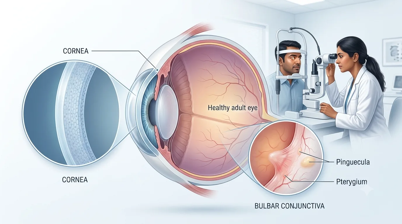
When you look at your reflection the white you see around the colored part of your eye may be thought of as plain. In fact it is made up of a thin specialized membrane which we know as the bulbar conjunctiva. This see-through tissue which plays a key role in the eye’s surface also protects the eye and supports smooth eye movement.
Changes in that tissue also present signs of eye health. Symptoms like redness, swelling, visible blood vessels, pigmentation or a yellowish brown color may present in the conjunctiva. It is important to know what is normal and what requires medical attention which in turn will help you in taking better care of your eye health.
What Is the Bulbar Conjunctiva?
The bulbar conjunctiva is a thin transparent mucous membrane which covers the front of the sclera, what is also known as the white of the eye. It extends to the corneal edge or limbus which at the same time allows the visibility of the underlying sclera and blood vessels.
The conjunctiva is a continuous structure as a whole which in no way is a separate entity. It covers parts of the eye, folds into the upper and lower fornix and extends along the inner aspect of the eyelids. Microscopic study reports that conjunctival tissue has epithelial cells and also specialized cells that play a role in the maintenance of the ocular surface.
Because at all times the eyes are open the bulbar conjunctiva is exposed to air, allergens, dust and other environmental irritants.
Bulbar Conjunctiva and Palpebral Conjunctiva: What Is the Difference?
People out to research palpebral and bulbar conjunctiva may think they are terms for separate tissues. In fact they are different areas of the same continuous conjunctival membrane.
| Feature | Bulbar Conjunctiva | Palpebral Conjunctiva |
| Location | Covers the visible sclera | Lines the inner surface of the eyelids |
| Visibility | Easily seen when the eye is open | Usually seen when the eyelid is pulled down or everted |
| Movement | Relatively loose over parts of the sclera | More firmly attached to the eyelid |
| Common clinical changes | Redness, swelling, haemorrhage, pigmentation, growths | Papillae, follicles, inflammation and irritation |
Understanding the distinction between the bulbar conjunctiva and palpebral conjunctiva is of great value in ophthalmic exams in which some conditions present more clearly in one area as opposed to the other.
What Does the Bulbar Conjunctiva Do?
The bulbar conjunctiva plays a role in the health of the eye. It is a protective layer over the sclera and also is a component of the smooth interface between the eyelids and eyeball.
Conjunctival epithelium and goblet cells also play a role in maintaining the tear film environment. Mucin producing cells which are a part of this group also play a role in surface lubrication and in the even distribution of tears over the eye. Research which has been done on conjunctival and ocular surface cells supports the role of these specialized cells in the health of the eye surface.
In the tissue you will see a network of small blood vessels which are visible to the eye. They may dilate in cases of irritation or inflammation which is the reason that in many cases of conjunctival disease you will see noticeable redness.
What Is the Upper Bulbar Conjunctiva?
The term used for the upper section of the sclera which is below the upper lid is upper bulbar conjunctiva. Usually what you see of this area is minimal without pulling up the lid. When it comes to diagnosis of persistent irritation, inflammation, contact lens issues or certain eye surface diseases ophthalmologists will closely look at the superior conjunctival area.
For example, abnormalities affecting the superior limbal and conjunctival area have been documented in patients with superior limbic keratoconjunctivitis.
What Is Bulbar Congestion?
Bulbar congestion is a term used for when blood vessels in the bulbar conjunctiva become more visible and dilated. In layman’s terms this is also referred to as a red eye.
The redness presents with allergic or infectious conjunctivitis, dry eye related irritation, environmental exposure or other inflammatory conditions. In studies it has been reported that bulbar conjunctival hyperaemia is a sign of ocular surface inflammation. Allergic conjunctivitis in particular produces noticeable conjunctival hyperaemia.
Not at every red eye is it just conjunctivitis, in fact. We see in what way the redness is present and also note pain, light sensitivity, discharge or visual changes which will help the ophthalmologist in determining the cause.
Common Conditions Affecting the Bulbar Conjunctiva
Several conditions can change the appearance of this normally transparent tissue.
| Condition | Possible Appearance or Symptoms |
| Conjunctivitis | Redness, irritation, watering, itching or discharge |
| Subconjunctival haemorrhage | Bright red, sharply defined patch over the white of the eye |
| Chemosis | Puffy or swollen conjunctiva |
| Pinguecula | Localised yellowish or whitish raised area |
| Pterygium | Fibrovascular tissue growing from the conjunctiva towards the cornea |
| Conjunctival pigmentation | Brown, grey or darker areas on the conjunctival surface |
| Conjunctivochalasis | Loose conjunctival folds that may cause irritation, watering or dryness |
A subconjunctival hemorrhage is what happens when blood pools under the conjunctiva which in turn may produce a large red patch although at times it causes little pain. Studies report that it is a fairly common ophthalmic issue. Chemosis is a term we use for conjunctival swelling which results from extra fluid in the tissue. It may present in many inflammatory or systemic settings and should be evaluated when it is severe or persistent.
Pterygium involves fibrovascular growth originating from the bulbar conjunctival region, while pinguecula is another frequently encountered ocular-surface change.
What Does Muddy Conjunctiva Mean?
The term which we use sometimes is muddy conjunctiva which describes when the usually white part of the eye takes on a dull, brown or gray appearance. It is important to note that this is more of a descriptive term and not a stand alone diagnosis.
Pigment changes in the conjunctiva may be due to a number of factors. Some people are born with conjunctival pigmentation related to their skin type and also we have seen that there is a great deal of difference in pigmentation between populations.
Chronic irritation and allergic eye disease also present with changes in the appearance of the ocular surface. Thus if you develop a new muddy conjunctiva which also has associated itching, redness, discomfort or pigmentation that is changing in nature do not assume it is cosmetic. A new, enlarging, irregular or atypical pigmented conjunctival lesion requires an ophthalmic assessment to determine between harmless pigmentation and more serious conditions.
How Are Bulbar Conjunctival Problems Diagnosed?
An ophthalmologist will start out by inquiring into redness, itching, discharge, pain, eye rubbing, allergies, contact lens use, injury and the duration of symptoms. The eye is then examined with a slit lamp under high magnification. We see the distribution of redness, surface edema, pigmentation, growths and involvement of adjacent structures like the cornea and eyelids.
This distinction matters because similar-looking red eyes can have very different causes and treatments.
When Should You See an Eye Doctor?
Mild transient redness may present itself in the aftermath of dust exposure, long term use of screens or small scale irritation. As for the persistent or chronic bulbar congestion, that should be evaluated rather than treated at home with OTC eye drops.
Seek timely ophthalmic care if you notice redness which is accompanied by pain, light sensitivity, reduced or blurred vision, eye trauma or severe swelling. In contact lens users painful red eyes also require special care as contact lens wear is associated with serious corneal infections.
Avoid over the counter use of steroid eye drops which is advised by your ophthalmologist. We treat the root cause of the problem and not just the redness which you see.
Conclusion
The bulbar conjunctiva is at times very thin, but it is an important element of the eye’s protective and lubricating surface. We see that changes like consistent redness, bulbar congestion, swelling, atypical pigmentation or a muddy conjunctiva may be indicators of base ophthalmic-surface issues.
Most of what we see in terms of conjunctival changes can be brought up during a routine ophthalmic exam. If you are still red, irritated or uncomfortable in the eyes or you notice a new growth or change in pigmentation we at Vasan Eye Care can do an exam which will help us to determine the cause of the issue and if treatment is required.
Frequently Asked Questions
1. Is the bulbar conjunctiva the white part of the eye?
Not really. The white element which is the sclera. The bulbar conjunctiva is a thin transparent membrane which covers the front of the sclera. As it is transparent this is what allows the white of the sclera and also the small conjunctival blood vessels to be seen.
2. What is the difference between palpebral and bulbar conjunctiva?
The palpebral and bulbar conjunctiva make up one continuous membrane. The palpebral which lines the inner eyelids, and the bulbar which covers the visible sclera and which also goes to the edge of the cornea.
3. What causes bulbar conjunctiva redness?
Redness is a sign which presents when the conjunctival blood vessels expand. Causes may include allergies, infection, dryness, environmental irritation and also other inflammatory eye conditions. Persistent redness in particular with pain, photophobia or visual changes requires professional assessment.
4. Is bulbar congestion dangerous?
Bulbar congestion is a term which is used to describe a look rather than a diagnosis. Many of the causes are minor in nature, but some eye issues may cause great redness. Pain, light sensitivity, reduced vision, trauma or contact-lens associated redness should not be ignored.
5. Is muddy conjunctiva a disease?
No. Muddy conjunctiva is a term used to describe conjunctiva which appears dull, brownish or pigmented instead of clear. This is a wide indication which may include many causes so new or different pigmentation should be looked at in detail instead of diagnosis which is based only on color.