
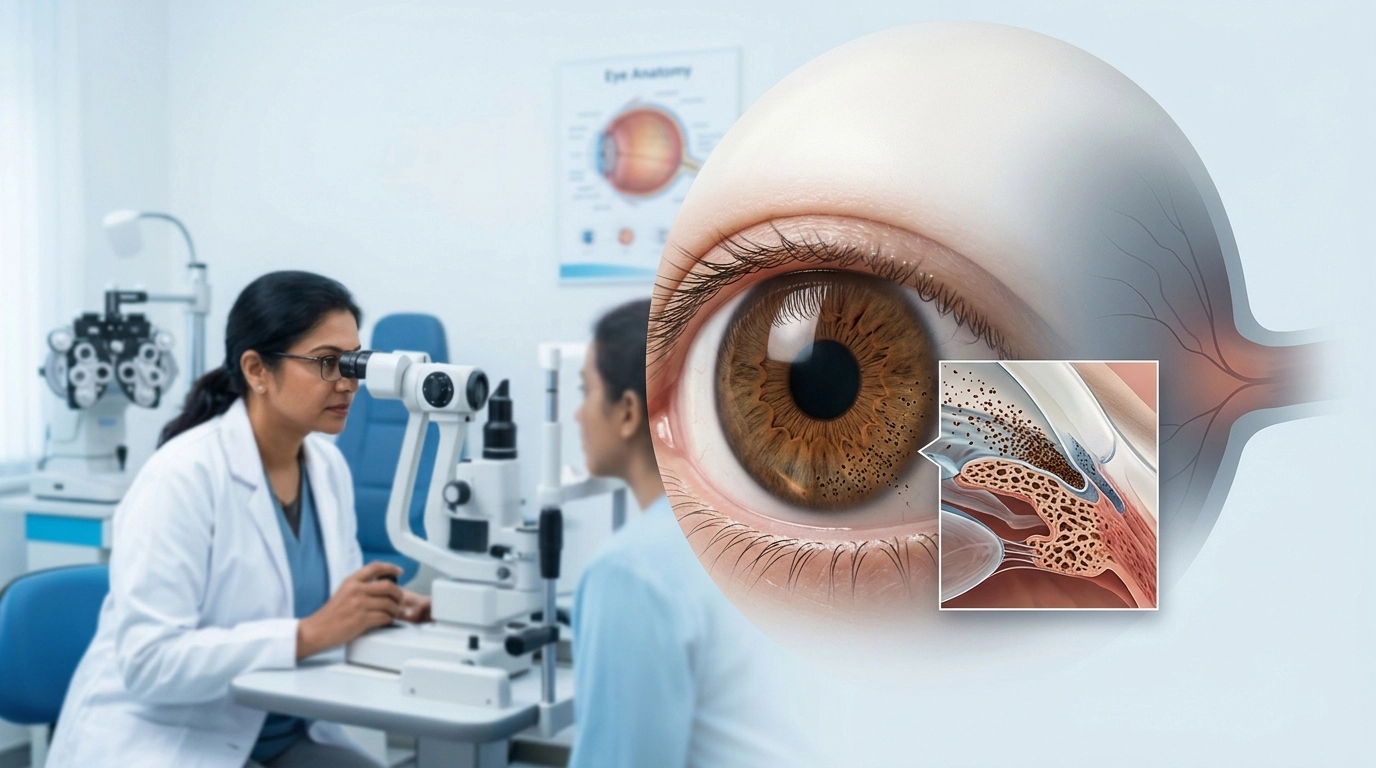
Glaucoma is a disease that mostly affects the elderly but also has an affinity for younger adults. Pigmentary glaucoma is a type which presents in young people. It is a result of pigments from the iris which get into the eye’s drain system. Over a period of time that pigment may block the natural outflow of fluid from the eye which in turn causes the high eye pressure to go up and damage the optic nerve.
What we see is that at the early stages many people do not notice any change in their eyesight. Between pigment dispersion and glaucoma, we often see that patients don’t recognize which is which also, which is why it is important to attend regular eye exams and to control eye pressure when it is required all of which play a role in reducing the risk of permanent vision loss.
What Is Pigmentary Glaucoma?
Pigmentary glaucoma which is a form of secondary open angle glaucoma. It is also related to a condition that we call pigment dispersion syndrome.
The iris which is the colored element of the eye. It’s at the back where we find pigments. In pigment dispersion syndrome very small particles of this pigment are released and they will travel through the eye’s interior front section.
Some particles which do end up in the trabecular meshwork, the tissue that is in charge of fluid outflow from the eye, do also play a role. Should large amounts of pigments be affected by this it may not be able to drain fluid out of the eye properly. That in turn may cause an increase in intraocular pressure or IOP.
Some particles which do end up in the trabecular meshwork, the tissue that is in charge of fluid outflow from the eye, do also play a role. Should large amounts of Pigmentary glaucoma be affected by this it may not be able to drain fluid out of the eye properly. That in turn may cause an increase in intraocular pressure or IOP
| Condition | What Happens? |
| Pigment dispersion syndrome | Pigment is released from the back of the iris and becomes deposited in different structures within the eye. Eye pressure may be normal or elevated. |
| Pigmentary glaucoma | Pigment interferes with fluid drainage, contributing to raised eye pressure and glaucomatous optic nerve damage. |
What Causes Pigmentary Glaucoma?
Pigmentary glaucoma may present with a wide variation in it’s physical features between individuals. But in general, the disease appears as a result of the iris’ rear surface chronically touching or pressure up against the internal eye structures which cause pigments to break off and clog fluid outflow.
In certain people, which includes those with a large anterior chamber and a concave iris, the movement of the iris may cause friction on the structures which support the lens. This friction results in the release of microscopic pigment particles.
The particles which pass through the aqueous humour, that is the clear fluid in the front of the eye, and may deposit in the drainage angle. As the trabecular meshwork becomes very pigmented or its function is impaired drainage resistance increases.
Also it goes that high eye pressure may develop. With time if the increase is continuous or repeated the optic nerve may get damaged.
Who Is More Likely to Develop Pigmentary Glaucoma?
Pigment dispersion syndrome and pigmentary glaucoma present in younger and middle aged adults more so than many other types of glaucoma. Also these conditions have traditionally been reported to affect more people with myopia or short sightedness.
However, myopia is a hereditary condition which may or may not develop in some individuals. Also it has been seen that pigment dispersion does not in all cases lead to glaucoma.
An ophthalmologist looks at many variables which include eye pressure, optic nerve appearance, pigment in the eye, drainage angle and visual field results.
Regular follow up is especially important once pigment dispersion is diagnosed as glaucoma tends to develop slowly without clear warning signs.
What Are the Symptoms of Pigmentary Glaucoma?
Many of the early stages of pigmentary glaucoma go unnoticed by many patients. That is why we see to it that we get in the habit of having our eyes checked at regular intervals.
Some may have transient symptoms as eye pressure goes up. These may present as dimmed vision, seeing colors or rainbows around light, eye discomfort, headache or redness. Symptoms also may present after vigorous physical activity or at other times of increased pigment release.
However, symptoms in and of themselves do not confirm the disease.
As glaucoma progresses, peripheral or side vision may gradually fall out. Also this may play out over a long time which in turn causes the brain to adapt and a person may not notice the loss until the damage is great.
Sudden severe eye pain, large vision loss, nausea or continuous halos should be evaluated right away as many eye conditions present with these symptoms.
How Is Pigmentary Glaucoma Diagnosed?
A full assessment for glaucoma is required to properly pigmentary glaucoma diagnosis.The ophthalmologist looks for signs of pigment dispersion and also reports on the effect is probably a typographical error and should be an “effect” of glaucoma on the optic nerve.
| Eye Test | Why It May Be Performed |
| Eye pressure measurement | Measures intraocular pressure and identifies elevated or fluctuating pressure. |
| Slit-lamp examination | Allows the ophthalmologist to look for characteristic pigment deposits and iris changes. |
| Gonioscopy | Examines the eye’s drainage angle and the amount of pigment present in the trabecular meshwork. |
| Optic nerve examination/OCT | Assesses the optic nerve and retinal nerve fibre layer for glaucomatous damage. |
| Visual-field test | Checks whether glaucoma has caused areas of peripheral vision loss. |
| Corneal thickness measurement | Helps the ophthalmologist interpret eye-pressure measurements more accurately. |
One which is true is a vertical pigmentation on the inner corneal surface which is called a Krukenberg spindle. Also in some patients you may see spoke-like areas where pigment has been lost from the iris.
These indicators are for diagnosis but there is no one tell-tale sign for pigmentary glaucoma. Eye pressure, drainage angle look, optic nerve health and visual field studies must be looked at together.
How Is Pigmentary Glaucoma Treated?
In pigmentary glaucoma treatment we aim to lower eye pressure which in turn reduces the risk of further optic nerve damage.
Treatment is tailored to the patient’s pressure levels, optic nerve findings, visual field results, age and rate of disease progression.
Eye Drops
Prescriptive glaucoma eye drops are a main treatment for reducing intraocular pressure. By which the eye in question may produce less fluid, or better drain what it does produce.
Patients must take their medication as prescribed. Glaucoma treatment does better in terms of control rather than permanent cure, if a patient stops their meds they will risk increased eye pressure.
Laser Treatment
Laser treatments are a choice for some patients.
In the case of Selective Laser Trabeculoplasty which is also known as SLT we target the trabecular meshwork and see to it that outflow of fluid improves. As pigmentary glaucoma may present with a very pigmented drain angle the ophthalmologist does a careful evaluation to determine if laser treatment is right and what the best approach should be.
Laser treatments are a choice for some patients. In the case of Selective Laser Trabeculoplasty which is also known as SLT we target the trabecular meshwork and see to it that outflow of fluid improves. As pigmentary glaucoma may present with a very pigmented drain angle the ophthalmologist does a careful evaluation to determine if laser treatment is right and what the best approach should be.
Glaucoma Surgery
When medication and laser options do not sufficiently reduce the pressure, surgery may be recommended.
Depending on the degree of glaucoma and the patient’s eye anatomy, an ophthalmologist may choose conventional glaucoma surgery, drainage implants or some minimally invasive glaucoma procedures.
The aim is to develop and improve passages which will allow fluid out of the eye and in that way reduce pressure.
Can Pigmentary Glaucoma Cause Blindness?
Untreated or poorly managed pigmentary glaucoma may result in long term optic nerve injury that is permanent and loss of vision that is reversible. The extent of which is determined by the level of intraocular pressure increase, the time the pressure has been uncontrolled and how quickly the disease progresses.
At the same time it is a positive that early diagnosis and proper treatment may greatly improve results for preservation of useful vision.
Vision does not usually return once it is lost from glaucoma which is why early detection is so important.
Living With Pigmentary Glaucoma
Being diagnosed with glaucoma is not a ticket to severe vision loss. Many patients report useful vision for years after diagnosis if the disease is identified early and managed well.
Patients must report to their ophthalmologist all the medications they are on including steroid eye drops, tablets, inhalers or skin preparations as steroids may raise eye pressure in some people.
Conclusion
Pigmentary glaucoma is a result of pigment from the iris which causes problems with fluid outflow in the eye and in turn causes damage to the optic nerve. Also at early stages it may not present with any noticeable symptoms and hence by the time a person reports visual changes the diagnosis may already be delayed.
An in depth evaluation which includes measurement of intraocular pressure, gonioscopy, optic nerve evaluation, OCT and visual field testing is a tool to diagnose pigment dispersion syndrome as opposed to glaucoma.
When appropriate we may use glaucoma medications, laser treatments or surgery which do help in the control of eye pressure and also in preserving what vision is left. If you have been diagnosed with pigment dispersion, high eye pressure or in fact have a family history of glaucoma we recommend that you have regular eye exams with our ophthalmologists at Vasan Eye Care which in turn may help in early identification of changes and in turn guide what is the best care.
Frequently Asked Questions
- Is pigment dispersion syndrome the same as pigmentary glaucoma?
No. Pigment dispersion syndrome is a condition which has pigment from the back of the iris being released and deposited in the eye. Pigmentary glaucoma is a diagnosis given when the condition is associated with glaucomatous damage to the optic nerve, also usually we see raised or fluctuating intraocular pressure.
- What age does pigmentary glaucoma usually occur?
It affects young and middle aged adults and also tends to present itself before many other types of glaucoma. But age is not the only determinant of risk. Any person who has suspicious eye pressure readings, optic nerve issues or pigment dispersion should follow the exam regime which is put forth by an ophthalmologist.
- Can exercise increase eye pressure in pigmentary glaucoma?
Some patients with pigment dispersion may have pigment release and increased eye pressure during or after intense exercise. This is not to say that all patients should forgo exercise. Speak to your ophthalmologist about your exercise routine instead of stopping at the drop of a hat.
- Can pigmentary glaucoma be cured permanently?
There is at present no cure for which we have success in the reversal of glaucomatous optic nerve damage. But in the case of pigmentary glaucoma treatment we are able to lower eye pressure which in turn greatly reduces the risk of additional damage. It is out that patients do require long term follow up even when their pressure is well managed.
- How often should someone with pigment dispersion have an eye examination?
Follow up frequency is determined by eye pressure, optic nerve appearance, visual field results and other risk factors. In the case of stable pigment dispersion some may require less frequent monitoring than that of a person with established glaucoma. Your ophthalmologist will put together a schedule of exams based on your individual risk.