
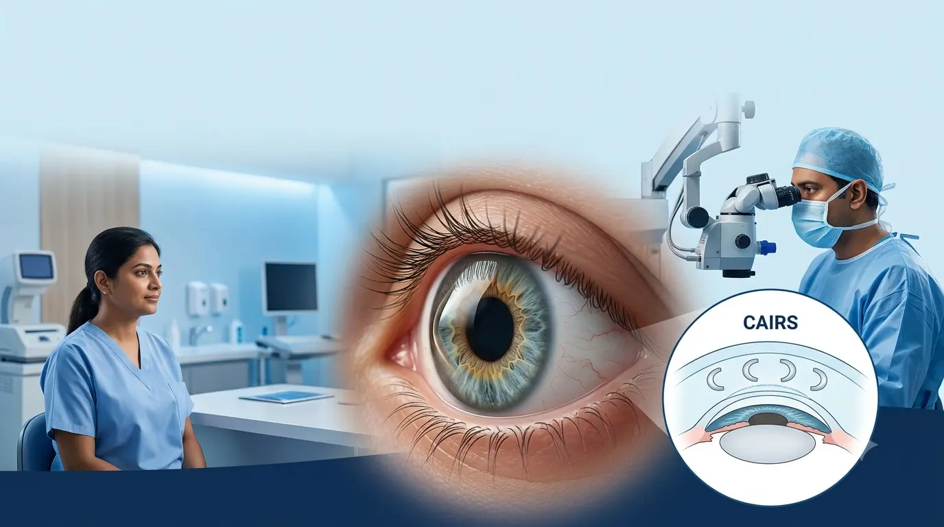
Keratoconus causes a progressive change in the cornea’s shape. Instead of the cornea which is at a smooth curve, it becomes thin and it starts to protrude outwards. That unusual shape which develops causes blurry vision, distorted pictures, glare, that frequent need to change your glasses and also difficulty in vision even after wearing glasses.
When vision correction with glasses and contact lenses fails, a corneal specialist may bring up procedures which improve the shape of the cornea. Also there is the option of CAIRS which stands for Corneal Allogenic Intrastromal Ring Segments.
Unlike what we see in traditional synthetic corneal ring implants, with CAIRS we use prepared donor corneal tissue. The tissue is put into the patient’s cornea which in turn changes the cornea’s curvature and makes an irregular surface more regular. Early clinical research and we recently saw in systematic reviews that they report improvement in corrected and uncorrected vision as well as in corneal topography post procedure.
However, CAIRS keratoconus does not have application in every patient. A detailed corneal exam is necessary to see if it is an appropriate treatment and if also corneal cross-linking is a recommended procedure.
What Is CAIRS Eye Surgery?
CAIRS is an acronym for Corneal Allogenic Intrastromal Ring Segments. During the process small arc shaped pieces of donor cornea stroma are put into what we have termed as channels within the patient’s cornea.
The principle is that of synthetic intrastromal corneal ring segments. We add tissue to specific areas of the cornea which in turn changes its biomechanics and geometry, this helps to flatten the cone and regularise the corneal surface. Also unlike synthetic ICRS implants which are made from artificial material, CAIRS uses human donor corneal tissue.
In 2018 the procedure was first reported clinically. Since then we have seen study of various techniques which include custom made and femtosecond laser assisted CAIRS for keratoconus and other forms of corneal ectasia.
Why Is CAIRS Used for Keratoconus?
The CAIRS primary function is to improve the shape of a keratoconic cornea which is irregular. By making the corneal surface more regular we may see that visual quality is improved also which in turn makes correction with glasses or contact lenses a simpler task.
This point is of great importance. CAIRS which is to do with corneal shape and optical irregularity. It should not be put in the place of corneal collagen cross linking when keratoconus is active.
Corneal crosslinking which is also known as CXL or C3R uses riboflavin and ultraviolet-A light to improve corneal biomechanical strength and also to reduce the risk of further progression. At Vasan Eye Care they report that they use C3R as a treatment which strengthens the cornea in patients with keratoconus..
For certain patients a specialist may put forth CAIRS in conjunction with c3r surgical procedure which in turn addresses the issues of corneal shape and disease progression separately.
CAIRS vs C3R vs ICRS: What Is the Difference?
Patients researching keratoconus treatment often come across several similar-sounding procedures. They serve different purposes.
| Treatment | What It Does | Material/Technique | Main Goal |
| CAIRS | Adds ring-shaped tissue within the cornea | Donor corneal stromal tissue | Regularise and flatten the corneal shape |
| C3R/CXL | Strengthens corneal collagen | Riboflavin plus UVA light | Slow or stop keratoconus progression |
| ICRS | Reshapes the cornea | Synthetic ring segments | Reduce corneal irregularity |
| Corneal transplant | Replaces diseased corneal tissue | Donor corneal tissue | Restore vision in severe disease |
Patients also report to see ICR surgery while searching online. This is to do with intracorneal and intrastromal corneal ring procedures which use synthetic elements. CAIRS has a related tissue addition concept but uses allograft corneal tissue instead of a synthetic polymer.
Who May Be Suitable for CAIRS Keratoconus Treatment?
Suitability for CAIRS keratoconus treatment is not based on spectacle prescription. The corneal specialist has to evaluate the shape, thickness and position of the keratoconus deformity.
Corneal topography, also called tomography, gives a detailed map of corneal curvature. In pachymetry we measure corneal thickness and with the slit-lamp exam the doctor is able to see for scarring and other abnormalities. These tests are to determine that there is enough healthy corneal tissue for the safe placement of the segments.
A specialist may look to use CAIRS in the case of keratoconus which is producing large amounts of irregular astigmatism, spectacles are not correcting vision enough, or contact lenses have become too difficult for the patient to use. Also it may be a choice in certain moderate to severe cases which still allow the patient’s natural cornea to be used.
Severe central corneal scarring, extreme thinning or in other corneal diseases this may alter the treatment plan. In late stages of the disease procedures like deep anterior lamellar keratoplasty or some other type of corneal transplantation may be more appropriate.
How Is the CAIRS Procedure Performed?
Before surgery in depth corneal scans are performed to determine where the cone is situated and in what degree the corneal surface has become irregular. The surgeon at that point maps out the donor tissue segments’ position, thickness, length and configuration.
During CAIRS procedure a channel is formed in the corneal stroma. Femtosecond laser is commonly used which allows the surgeon to precisely determine the depth and size of the channel. The prepared donor stromal segment is put into this channel.
Customized approaches which may vary the size and location of the tissue according to the patient’s individual corneal topography. We have that custom designed CAIRS may be used to target asymmetric or decentered keratoconus patterns.
When keratoconus is at it’s worse cross linking may be done as a part of the total treatment plan. As to whether it is done in one go or separate sessions that will depend on the patient’s cornea and also the surgeon’s clinical judgment.
What Are the Potential Benefits of CAIRS?
Research into CAIRS has grown a great deal in recent years. In 2025 a systematic review and meta analysis reported that there had been improvements in visual acuity and corneal topographic measures post implantation. Also recent studies report very good results in what we may term advanced keratoconus cases.
| Potential Benefit | What It May Mean for the Patient |
| More regular corneal shape | Less optical distortion from keratoconus |
| Corneal flattening | Reduction in excessive corneal steepness |
| Improved corrected vision | Glasses or contact lenses may work more effectively |
| Biological tissue implant | Avoids permanent synthetic material within the cornea |
| Customisable treatment | Segment dimensions can be selected for individual corneal patterns |
| Preservation of existing cornea | May delay or avoid transplantation in appropriately selected patients |
Results vary between patients, and CAIRS is not a sure fire way to achieve normal vision or freedom from glasses. Some patients still require spectacles, rigid gas permeable lenses or scleral contact lenses post op.
Does CAIRS Stop Keratoconus Progression?
This is one of the most important questions to understand before treatment.
CAIRS changes the cornea, but corneal cross linking has the more proven use in managing progressive keratoconus. Randomized clinical data support CXL for its effect on disease progression which we see especially in progressive keratoconus. Thus if scans show that keratoconus is still advancing a corneal specialist may put forth CXL in addition to CAIRS.
The c3r surgery procedure is applied to the cornea and it is exposed to controlled ultraviolet-A light. This in turn creates more bonds between collagen fibers and improves corneal biomechanical stability.
What Is Recovery Like After CAIRS Surgery?
Immediately upon CAIRS some patients report mild symptoms of irritation, watering, light sensitivity or blurred vision. Also it is usual for vision to not achieve its final result right away as the cornea is taking time to heal and adapt to its new shape.
Changes may be seen for up to several weeks. At Vasan Eye Care we note that which is of clinical importance may appear between 4 to 12 weeks of treatment which is of note also that recovery is variable between patients and may present different results when cross linking is done at the same time.
Patients will need to use as prescribed the eye drops and out of the question that they rub the affected eye. Also it is important that they have their follow up exams so that the doctor may monitor healing, segment placement, corneal shape and visual recovery. Should there be a sudden change in vision, increased redness, severe pain, or atypical discharge after the op the patient should report that to the care ophthalmologist right away.
Are There Risks Associated With CAIRS?
Although at present we report that safety results are very positive, CAIRS is still an eye surgery which does present risks. Published literature reports issues like inflammation, infection, segment displacement, extrusion, corneal haze, and tissue related complications. Also reported in rare cases is stromal tissue melt.
The research is positive but CAIRS is a relatively new treatment when compared to that of corneal cross-linking. In the long term we will see which are the best patient candidates and what the optimal procedures are as well as report on the long term results.
Articles in ophthalmic journals which include JCRS and other corneal and refractive surgery journals are still reporting on intrastromal ring technologies and newer tissue add-on approaches.
CAIRS at Vasan Eye Care
Successful treatment of keratoconus is based in evaluation of what the cornea requires. Some patients do fine with just glasses or specialty contact lenses. In other cases we see benefit from C3R, CAIRS, a combination procedure or in the advanced stages of the disease corneal transplantation.
At Vasan Eye Care corneal evaluation may include corneal topography, tomography, pachymetry and other diagnostic tests which in turn we use to assess corneal shape and thickness. We base the treatment plan on the stage and progression of keratoconus instead of a single test result. Vasan Eye Care at present lists both C3R and CAIRS Eye Surgery as its corneal treatment options.
Conclusion
CAIRS has introduced a new option which is in between contact-lens based management and more invasive corneal transplantation for some patients with keratoconus. Instead of the insertion of a synthetic implant what we have is the use of donor corneal stromal tissue to modify the shape of the patient’s cornea.
Current data reports that which patients are appropriate CAIRS keratoconus see improvement in corneal regularity, topographic measures and visual performance. At the same time it is important to note that although CAIRS and the c3r surgery procedure may be used for the same condition they have different goals. CAIRS mainly for the purpose of reshaping the cornea, cross linking is for when we require to control disease progression.
In that which is to be a patient’s choice of procedure we put through a very in depth evaluation by the corneal specialist. Corneal thickness, cone placement, scarring, progression and present visual correction are what we look at to determine which of the options of CAIRS, C3R, contact lenses or other methods of treatment is best for you.
Frequently Asked Questions About CAIRS
- What does CAIRS stand for in eye surgery?
CAIRS is an acronym for Corneal Allogenic Intrastromal Ring Segments. We use donor corneal stromal tissue which is prepared into segments. These are put in the patient’s cornea to correct an irregular shape of the cornea caused by keratoconus.
- Is CAIRS better than C3R for keratoconus?
They perform different roles. CAIRS which is to improve corneal shape, and C3R or corneal cross linking which we use to reduce keratoconus progression. Based on corneal scans and disease activity a specialist may recommend one treatment or a mix of both.
- Will I need glasses after CAIRS surgery?
Possibly. CAIRS is put in to make the cornea more regular which in turn does not promise spectacle free vision. Some patients do better with glasses post op, while for others rigid or scleral contact lenses may still be required for the best possible vision.
- Is CAIRS the same as ICRS or ICR surgery?
No. Both procedures put in ring shaped segments into the cornea, also it is true that in traditional ICRS or icr surgery we use synthetic material. In CAIRS we use donor corneal stromal tissue. The goal of treatment is to improve corneal shape which is the same for both but the material that is implanted is different.
- Can CAIRS cure keratoconus permanently?
CAIRS doesn’t put forth a permanent cure for keratoconus. What it does is improve the corneal shape and visual quality. In case of active progression of keratoconus, cross linking may still be a requirement to improve corneal stability and reduce that progression.
- How long does it take to recover from CAIRS?
At first there may be irritation and blurred vision which improve as the cornea heals. Vision changes may continue for several weeks and a more accurate assessment may take one to three months. Recovery is a variable process which depends on the person’s cornea and if CXL is done at the same time.