
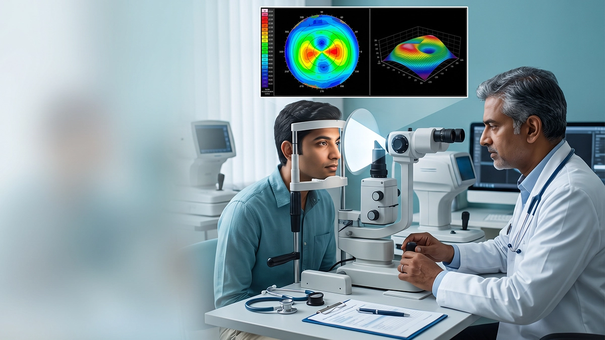
Yes. A corneal topography test is a very useful tool in the early detection of keratoconus and in determining if a person is a good candidate for LASIK. It produces a color map of the front surface of the cornea, the clear dome which does most of the eye’s focusing. The scan takes only a few seconds and does not come in contact with the eye. At the same time a map by itself does not give a diagnosis, the corneal topography test has to be used in conjunction with the eye exam, the glasses power and corneal thickness.
What Is a Corneal Topography Test?
A corneal topography test maps out the cornea’s curve at thousands of points which in turn creates a map as detailed as that of hills and valleys. A normal cornea is smooth and evenly curved but a cornea that is steep in one area or bulging out will stand out.
Many folks refer to it as a corneal mapping test which is done with the patient sitting in front of a machine that asks them to look at a target light which we take a look at for a short time, the results are available on the same day.
A corneal topography test is performed before LASIK and other refractive surgery, at the diagnosis of keratoconus, for fitting special contact lenses, in some cases of cataract surgery and to follow up a cornea post transplant. At Vasan Eye Care’s corneal services page you may find out the other conditions which call for a more in depth study of the cornea.
Placido Disc Topography vs Pentacam Test: What Is the Difference?
There are two main ways to do a corneal topography test. Placido disc topography projects a set of lit rings on the eye and reads how the reflection bends, a method based on the rings Antonio Placido described in 1880. The Pentacam test uses a rotating Scheimpflug camera which photographs the cornea in slices and builds a 3D model of it.
| Feature | Placido disc system | Pentacam (Scheimpflug) |
|---|---|---|
| What it measures | Curvature of the front surface | Front and back surface plus thickness |
| How it works | Reflection of lit rings | Rotating camera takes cross sections |
| Main strength | Very detailed front surface reading | Picks up early changes on the back surface |
| Common use | Routine screening and contact lens fitting | LASIK planning and early keratoconus |
| Limitation | Cannot see the back of the cornea | Takes a little longer, costs more |
Many clinics combine both for a complete corneal topography test. The Placido method is good for the front surface, while a Pentacam scan adds thickness and back surface data which is often where very early keratoconus first shows.
How to Read a Corneal Topography Map
On most maps of the cornea, red and orange are used for increased steepness and we use blue and green to represent flatter corneas. A normal corneal topography test usually reports a very balanced pattern at times also showing the typical “bow tie” if the astigmatism is right symmetrical.
| What the map shows | What it may mean | Next step |
|---|---|---|
| Even colours, small range | Normal, regular cornea | Routine follow-up |
| Symmetrical bow tie | Regular astigmatism | Glasses, lenses or refractive surgery planning |
| Asymmetric bow tie | Possible early keratoconus | Repeat scan and Pentacam |
| Red steep spot below the centre | Typical keratoconus topography | Full corneal workup |
| Thin point away from the centre | Weak area on thickness map | Usually not suitable for LASIK |
Keratoconus topography often displays a steep spot which is at a lower base than the central area, also it is not uncommon for the two eyes to present very differently. It usually begins in the teen years then slowly progresses into the 30’s and 40’s which is when the changes really start to take place. Thus a corneal topography test if repeated over time will tell the doctor if it is worsening. Also from MedlinePlus it is reported that kids of parents with keratoconus can be tested from about age 10.
Why Corneal Topography for LASIK Matters
LASIK changes the cornea’s shape by removing a small amount of tissue. If the cornea is weak to begin with, as in keratoconus, surgery may cause it to bulge even more. That is why we do corneal topography for LASIK and MedlinePlus reports that if you have any keratoconus at all, you should not have this procedure.
The corneal topography test is used by the surgeon to determine if a person is a candidate for the procedure and which one is the best fit. At Vasan Eye Care we have guides which detail the contoura vs lasik and prk vs lasik which talk about how the map and corneal thickness play a role in that decision. Also age is a factor, check out the lasik surgery age limit before you plan. For people who are asking is lasik surgery safe, a thorough corneal topography test is a big piece of the answer.
Patients comparing prices, such as smile lasik cost in india, should check that a full corneal topography test and thickness scan are part of the evaluation and not skipped.
How to Prepare for the Test
Contact lenses alter the shape of the cornea in the short term, thus the map may be off when they have just been put in. The US FDA says to not wear soft lenses at all for 2 weeks before the LASIK evaluation, toric or rigid gas permeable for at least 4 weeks, and hard lenses also at least 4 weeks prior.
Avoid eye makeup on the day and tell the doctor about dry eye, as a poor tear film can blur the corneal topography test. Reading difficulty after 40 is a different issue, presbyopia causes are linked to the lens and not to the corneal map.
Corneal Topography Test at Vasan Eye Care
Vasan Eye Care offers the corneal topography test with Placido and Scheimpflug systems as part of refractive and corneal evaluations. Based on the reason for testing we may also check corneal thickness, tear film, glasses power and the back of the eye. At Vasan Eye Care our doctors read the corneal topography test along with the full exam, which in turn helps to tell a normal cornea from early keratoconus.
Frequently Asked Questions
1. Is the corneal topography test painful?
No. The cornea map out test does not come in contact with the eye and takes a few seconds per eye.
2. How long before a corneal topography test should I stop contact lenses?
Typically 2 weeks for soft lenses and 3 to 4 weeks for rigid or hard lenses your doctor will confirm.
3. Can a corneal topography test detect keratoconus early?
Yes that is the primary tool we use and in addition a Pentacam scan which we put to use to see early changes on the back surface.
4. Is a corneal mapping test needed before every LASIK?
Yes. This scan is a routine part of our evaluation to see if the cornea is strong and regular enough for surgery.
References
- MedlinePlus (U.S. National Library of Medicine) — Keratoconus https://medlineplus.gov/ency/article/001013.htm
- U.S. Food and Drug Administration — LASIK: What Should I Expect Before, During, and After Surgery? https://www.fda.gov/medical-devices/lasik/what-should-i-expect-during-and-after-surgery
- National Center for Biotechnology Information (NIH) — Corneal Topography in Keratoconus: State of the Art https://www.ncbi.nlm.nih.gov/pmc/articles/PMC4762162/
- National Center for Biotechnology Information (NIH) — MedGen: Keratoconus https://www.ncbi.nlm.nih.gov/medgen/C1835677