
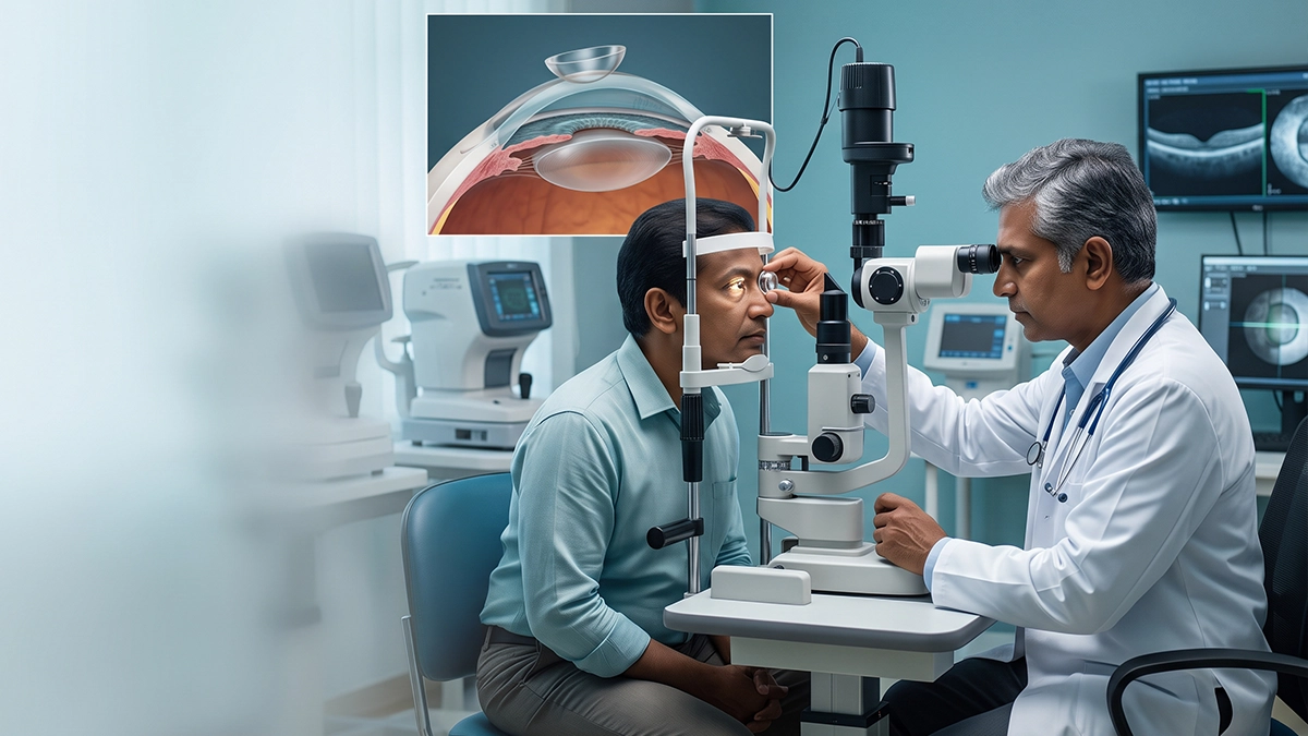
A gonioscopy test for glaucoma examines the drainage angle at the point where the cornea and iris meet. This area is responsible for the exit of aqueous humour from the eye. The test allows an ophthalmologist to determine if the angle is open, narrow or closed and also to identify which forms of glaucoma require different treatments. It is particularly important when narrow angles or angle-closure glaucoma are suspected.
What Is a Gonioscopy Test for Glaucoma?
In the gonioscopy procedure vasan eye care uses a special contact lens which they call a gonioscope with a slit-lamp microscope. The lens allows ophthalmologists to see the anterior chamber angle which in routine exams cannot be seen because of the way light reflects in the cornea.
This angle test for glaucoma lets the ophthalmologist use it to look at structures which include the trabecular meshwork, scleral spur and iris insertion. The trabecular meshwork is a key element in the eye’s natural fluid outflow. In greater detail a Vasan Eye Care guide goes into the full role of gonioscopy in the assessment of the eye’s drainage systems.
Why Is Gonioscopy Important in Glaucoma?
A gonioscopy test for glaucoma helps distinguish open angle glaucoma from narrow angle or acute closure glaucoma. This is important as treatment options and emergency risk factors are different between these groups. In narrow angle glaucoma test the iris is very near the trabecular meshwork. Should the angle close up, outflow of aqueous humor may be impeded and intraocular pressure can rise.
According to the American Academy of Ophthalmology’s guidelines on gonioscopy which is a procedure for the assessment of the anterior chamber angle and is an element of glaucoma evaluation. Glaucoma is a large health issue in India. National Blindness Survey which ran from 2015 19 reported that glaucoma caused visual impairment in 59.6 per 10,000 adults aged 50 years or over. The study which included 85,135 participants reported that. Also in 2026 National Blindness Survey reported glaucoma related blindness in 10.4 per 10,000 adults aged 50 years or over.
| Gonioscopy finding | What it means | Why it matters |
| Open angle | Drainage structures remain visible | Supports assessment for open-angle glaucoma |
| Narrow angle | Iris lies close to the drainage pathway | Indicates increased risk of angle closure |
| Appositional closure | Iris temporarily contacts the trabecular meshwork | May open during indentation gonioscopy |
| Synechial closure | Adhesions keep part of the angle closed | Suggests more persistent structural closure |
How Is the Gonioscopy Procedure Performed?
During the course of a gonioscopy eye test which is performed by an ophthalmologist see that usually anesthetic drops are put in the eye. The patient sits at the slit lamp as a gonioscopy lens gently touches the corneal surface.
The doctor at this point looks at various elements of the drainage angle. A typical gonioscopy test for glaucoma is of short duration to that of a full eye exam. Temporary mild blur may present from the exam gel or anesthetic used.
In dynamic or indentation gonioscopy gentle pressure is applied which in turn helps to determine if a closed angle can open. This in turn is useful in telling between temporary iris contact and permanent adhesions.
What Does the Anterior Chamber Angle Test Show?
The anterior chamber angle test does in fact look at more than just angle width. It also reports on abnormal pigmentation, new blood vessels, trauma which causes angle recession, inflammation and adhesions which are known as peripheral anterior synechiae.
| Finding | Possible clinical relevance | Additional evaluation |
| Narrow drainage angle | Risk of angle closure | Eye pressure, optic nerve and angle assessment |
| Heavy pigmentation | Pigment dispersion or other angle change | Glaucoma evaluation |
| Abnormal vessels | Possible neovascular process | Retinal and systemic assessment |
| Angle recession | Previous blunt eye trauma | Long-term glaucoma monitoring |
| Peripheral anterior synechiae | Permanent areas of angle closure | Assessment of glaucoma severity |
A gonioscopy test for glaucoma may therefore form part of a wider assessment that includes tonometry, optic nerve examination, OCT and visual-field testing.
Who May Need a Narrow Angle Glaucoma Test?
An ophthalmologist will put forward a narrow angle glaucoma test at the time of a routine exam which reveals a shallow anterior chamber or increased risk of angle closure. Also it may be a part of the work up for high eye pressure, glaucoma diagnosis or a history of acute angle closure.
People that have had to be tested for glaucoma may require to have a gonioscopy to properly classify the condition. The gonioscopy eye test also is used to assess patients with eye trauma, inflammation or atypical changes in the drainage angle.
Conditions involving altered anterior chamber anatomy may require additional assessment. Examples include malignant glaucoma and phacolytic glaucoma.
How Can Gonioscopy Results Affect Treatment?
Results of the angle test for glaucoma which in turn can be used to determine what treatments will be appropriate. In the case of a narrow or closed angle they may look at preventive or therapeutic laser treatment after a full clinical evaluation. For open angle disease vasan eye care may use pressure reducing medications, laser or surgery which will be based on the extent of the damage to the optic nerve and the pressure in the eye. In very advanced cases it may have to perform procedures such as the trabeculectomy procedure.
Inflammation also changes in the angle structure and drainage. Patients that present with eye pain, redness or light sensitivity may do well to be evaluated for conditions which include anterior uveitis signs.
The anterior chamber angle test in itself does not diagnose glaucoma. Ophthalmologists use it in conjunction with eye pressure, optic nerve appearance, OCT results and visual field studies.
When Is Urgent Eye Assessment Needed?
A gonioscopy test for glaucoma becomes particularly relevant when acute angle closure is a issue. Severe eye pain, a red eye, blurred vision, halos around lights, headache, nausea or vomiting may present with a sudden pressure rise. These symptoms require immediate ophthalmic evaluation. Delay in treatment during acute angle closure may put the optic nerve and vision at risk.
Eye Care at Vasan Eye Care
Vasan Eye Care which does glaucoma assessment of the patient’s symptoms, drainage angle anatomy, intraocular pressure and optic nerve atrophy. As per the exam which may also include use of the goniolens may do OCT, tonometry and visual field testing which in turn will be followed by discussion of what monitoring or treatment is appropriate.
Frequently Asked Questions
1. Is a gonioscopy test for glaucoma painful?
The test is usually pain free as anesthetic drops are used to numb the eye prior to the exam. There may be mild pressure from the lens or temporary irritation. If you do experience persistent pain after an eye exam, report it to an ophthalmologist.
2. What is the difference between gonioscopy and an eye-pressure test?
Tonometry measures eye pressure. Gonioscopy looks at the drainage angle which is responsible for aqueous humor outflow. While these tests present different info, a normal pressure reading doesn’t do away with the need for drainage angle evaluation.
3. Can a gonioscopy eye test detect narrow angles before symptoms begin?
Yes. A gonioscopy eye test which may identify narrowed or at risk angles before a patient shows any signs. Vasan eye care may see abnormal angle anatomy which in turn allows the ophthalmologist to evaluate the risk of future acute attacks and discuss what the best course of action is.
4. How often is an anterior chamber angle test needed?
There isn’t a one size fits all approach to testing. It depends on angle anatomy, glaucoma type, past results, eye procedures and other risk factors. An ophthalmologist is the best to determine when repeat gonioscopy is clinical.
References
- American Academy of Ophthalmology EyeWiki — Gonioscopy
https://eyewiki.aao.org/Gonioscopy - Glaucoma Blindness and Visual Impairment in India: Findings From the National Blindness Survey
https://pubmed.ncbi.nlm.nih.gov/42611182/